
Secure • Private • Science-backed
NAD vs peptides featured image showing peptide vials on a counter in dramatic lighting

NAD+ vs Peptides: The Differences, Explained

What You Should Know

  • NAD+ is not a peptide. They work through completely different mechanisms in the body.
  • Most longevity peptides (BPC-157, MOTS-c, GHK-Cu) have been studied mainly in animals, not humans. NAD+ has multiple human clinical studies behind it.
  • Some compounds frequently compared to NAD+ (MOTS-c, 5-amino-1MQ) actually depend on NAD+ to do their job, making them complementary rather than competing options.
  • You can measure your NAD+ levels with a blood test and confirm whether they change after treatment. You cannot do that with most peptides.

Walk into any longevity clinic or scroll through enough biohacking content, and you’ll find NAD+ and peptides listed in the same breath. Sermorelin. BPC-157. GHK-Cu. MOTS-c. NAD+ injections. They appear together on clinic menus, in wellness stacks, on podcasts. The implication is that they’re roughly the same kind of thing and you’re choosing between them.

They’re not the same kind of thing. NAD+ is a molecule your cells use to make energy. It’s inside every cell in your body right now. Peptides are short chains of amino acids that act like biological text messages, sending specific instructions to specific parts of the body. These two things work differently, have different amounts of research behind them, and solve different problems.

Grouping them together causes real confusion about what each does and when, or whether, either is worth pursuing. This article untangles the distinction and walks through six specific comparisons to help you think clearly about each one.

CLIA-Certified NAD Blood Test

Find out if your NAD+ is in the healthy range.
Jinfiniti Intracellular NAD Test on purple background

NAD+ Is Not a Peptide

The confusion makes sense. Both NAD+ and peptides come up constantly in anti-aging and longevity conversations, and some clinics sell them side by side. But they are chemically different things that do different jobs.

NAD+ (nicotinamide adenine dinucleotide) is a helper molecule your cells rely on to convert food into usable energy. Think of it as the fuel gauge for your cells. Every cell in your body needs it, all the time, to repair DNA, produce energy, and keep basic functions running. It is not built from amino acids and is not a peptide.

Peptides are different. They’re short chains of amino acids (the same building blocks that make up proteins) that carry targeted instructions. A peptide might tell your pituitary gland to release more growth hormone, or signal skin cells to produce more collagen. The scope is narrow by design. That is both their strength and their limitation.

As Eric Verdin documented in a 2015 review, NAD+ is involved in energy production, DNA repair, and activating proteins called sirtuins (which help regulate how cells age) in virtually every cell in the body. No single peptide does that. Each peptide has one job. NAD+ has hundreds.[1]

NAD+ also declines with age. By midlife, many adults have 40 to 50% less NAD+ than they did in their 20s. That drop tracks closely with the kind of fatigue, brain fog, and slower recovery that most people write off as just getting older. The good news is that NAD+ levels can be tested and raised.

How Peptides Work and Why the Evidence Varies

The word “peptide” just describes a molecular structure: a short chain of amino acids. It does not tell you what the peptide does. Your body makes thousands of them, and they serve completely different purposes. Insulin is a peptide. So are endorphins. The GLP-1 medications used for weight loss, like Ozempic, are also peptides, and they have some of the most rigorous human research of any drug in recent memory.

The peptides that come up in longevity clinics are a different category. Most of them are experimental. Many are sold as research compounds rather than regulated supplements, and their evidence comes almost entirely from animal studies, not human trials. That does not mean they do nothing. It means we do not yet know what they do in people, at what dose, or over what time frame.

The Regulatory Picture

Not all peptides have the same legal status, and that matters when you’re deciding what to put in your body.

Regulatory CategoryExamples
FDA-approved medicationsSemaglutide (Wegovy), tirzepatide (Zepbound)
Prescription-only compounded drugsSermorelin, ipamorelin, CJC-1295
FDA-flagged substances (safety concerns raised)BPC-157, MOTS-c
Unregulated research compounds5-amino-1MQ

BPC-157 and MOTS-c have both been flagged by the FDA for potential safety concerns, and both appear on WADA’s list of banned substances (the organization that oversees drug testing in competitive sports). This does not automatically make them dangerous. But it is information that most influencers leave out entirely.

NAD+ vs. Specific Peptides: A Side-by-Side Breakdown

Each peptide targets something different. The right question is not which one wins against NAD, but what problem you are actually trying to solve.

Here is an overview before the detailed comparisons:

CompoundWhat It IsWhat It TargetsHuman ResearchCan You Test It?
NAD+Energy molecule found in all cellsEnergy, cell repair, metabolism, cognitionMultiple controlled human trialsYes, via blood test
BPC-157Synthetic peptideTissue repair, gut healing, injury recoveryAlmost none (mainly animal studies)No
SermorelinSynthetic peptideGrowth hormone productionModerate (growth hormone studies)Yes, via IGF-1 blood test
GHK-CuNaturally occurring copper peptideSkin, collagen, wound healingModerate (mostly topical studies)No
MOTS-cPeptide from mitochondrial DNABlood sugar regulation, metabolismEarly stage (mainly animal studies)No
5-amino-1MQSmall synthetic moleculeSlowing NAD+ breakdown, fat metabolismAnimal studies onlyNo

BPC-157 vs. NAD+

BPC-157 is a synthetic peptide originally derived from a protein found in stomach juice. In animal studies, it has shown the ability to speed up healing in tendons, ligaments, and the gut lining, likely by encouraging the growth of new blood vessels and stimulating the cells that build connective tissue.[2]

The problem is that those results are almost entirely from animals. There are a handful of small human case reports, but no proper clinical trials. The FDA does not recognize BPC-157 as a legal supplement, and it cannot be sold as one.[3]

NAD+ does not repair a torn tendon the way BPC-157 is supposed to. What it does is keep the energy supply running in the cells doing the repair work. These are different tools:

  • BPC-157 may be worth a conversation with a doctor if you are dealing with a specific injury or chronic gut issues, with realistic expectations about the limited evidence
  • NAD+ is the stronger choice for energy, mental clarity, metabolic health, and overall cellular function, with human data to back it up

Sermorelin vs. NAD+

Sermorelin is a synthetic peptide that tells your pituitary gland (a small gland at the base of your brain) to release more growth hormone. Growth hormone naturally declines with age, which can contribute to muscle loss, fat gain, poor sleep, and slower recovery. Sermorelin works by mimicking the body’s own signal for growth hormone release rather than replacing the hormone directly.

The evidence for sermorelin is stronger than for most longevity peptides. Studies on growth hormone restoration in older adults have shown real improvements in muscle mass, body fat, and sleep quality. It requires a prescription and is administered through a compounding pharmacy.[4]

NAD+ and sermorelin work at different levels of the body:

  • Sermorelin works in the hormonal system, addressing growth hormone decline
  • NAD+ works inside individual cells, maintaining the energy supply that all hormones and repair processes depend on

Someone with both low and declining growth hormone might benefit from addressing both, but in the right order. They are not alternatives to each other.

GHK-Cu vs. NAD+

GHK-Cu is a small copper-containing peptide that naturally exists in your blood, saliva, and urine. Levels drop significantly as you age, and research has found that this decline is connected to things like thinning skin, slower wound healing, and hair loss. GHK-Cu works by activating the cells that produce collagen and elastin (the proteins that keep skin firm and elastic) and by helping regulate genes involved in tissue repair.

One analysis identified more than 4,000 human genes that GHK-Cu appears to influence. Among the peptides in this article, it has some of the more credible evidence, particularly for skin health.[5]

GHK-Cu and NAD+ are not competitors:

  • GHK-Cu targets structural repair in the skin and connective tissue
  • NAD+ targets energy production and cell maintenance across the entire body

Using both makes sense if both goals are relevant. They address different things.

MOTS-c vs. NAD+

MOTS-c is a small peptide produced inside your mitochondria (the parts of your cells that generate energy). Most peptides are encoded in the DNA inside your cell’s nucleus. MOTS-c is unusual because it comes from the DNA inside the mitochondria itself.

A 2015 study in Cell Metabolism found that it plays a role in regulating blood sugar and metabolism, and that it can mimic some of the metabolic benefits of exercise in animal models.[6]

MOTS-c works inside mitochondria. Mitochondria need NAD+ to function. The two are not alternatives. MOTS-c depends on adequate NAD+ to do its job. Low NAD+ undermines the very system MOTS-c is trying to support.

MOTS-c is also banned by WADA, has no published human clinical trials, and is sold as an unregulated research compound. For anyone interested in metabolic health, building a foundation with NAD+ first is the more sensible and evidence-backed approach.

For more context on how NAD+ compares to other mitochondrial health compounds, see Urolithin A vs. NAD+.

5-Amino-1MQ vs. NAD+

One clarification upfront: 5-amino-1MQ is not a peptide. It is a small synthetic molecule, and it shows up in longevity content because of its relationship to NAD+, not because it belongs in the same category as BPC-157 or sermorelin.

Here is what it does. Your body has an enzyme called NNMT that breaks down a building block your cells use to make NAD+. When NNMT is overactive, your NAD+ gets depleted faster than your body can replenish it. 5-amino-1MQ blocks that enzyme, slowing the breakdown.

In fat cell studies published on PubMed, it raised intracellular NAD+ levels by 1.2 to 1.6 times. Most interest has focused on fat loss and blood sugar regulation.[7]

There are no published human trials. It is sold as a research compound.

The relationship with NAD+ is synergistic:

  • 5-amino-1MQ slows how fast NAD+ gets used up
  • NAD+ precursors like NMN and NR increase how much NAD+ your body produces

These two approaches work on the same problem from different angles. Direct NAD+ supplementation has considerably more human research behind it.

Peptide Therapy vs. NAD Injections

Someone comparing peptide therapy to NAD+ injections is usually weighing two clinical options: a full peptide protocol from a longevity or regenerative medicine clinic, versus NAD+ delivered by injection (either directly into a vein or under the skin).

Peptide therapy protocols at clinics typically combine several different peptides at once. A common stack might include one peptide to raise growth hormone, another to help with injury recovery, and another to support skin or collagen. Each peptide delivers a targeted message to one specific system. That specificity is the appeal. But it also means that the protocol as a whole has almost never been studied, and the evidence for each individual component varies from moderate to essentially nonexistent.

NAD+ injections deliver one molecule that every cell in your body uses. The effect is broad rather than targeted. When your NAD+ levels are low, cells across multiple systems are running below capacity. An injection addresses that deficit everywhere at once, not just in one tissue or organ.

Peptide TherapyNAD+ Injections
How it worksEach peptide sends a signal to one specific systemRestores a molecule that all cells need to function
ScopeNarrow and targeted per peptideBroad effect across the whole body
ProtocolMultiple compounds, each with a different purposeOne molecule, one mechanism
Research qualityVaries widely; many compounds have no human trialsGrowing body of human clinical data
Can you test outcomes?Rarely, for most peptidesYes, via intracellular NAD+ blood test
Legal statusRanges from prescription to FDA-flaggedAdministered clinically; not FDA-approved as a drug

Peptide therapy protocols can run several hundred to over a thousand dollars a month depending on what is included. NAD+ injections carry their own clinic-dependent cost. Before committing to either, knowing your baseline NAD+ level tells you whether NAD+ deficiency is part of what you’re dealing with, which changes how any protocol should be sequenced.

For a closer look at what NAD+ injections involve clinically, see NAD injection side effects and what they mean.

How to Choose What to Address First

There is no single right answer, but starting with your goal, then looking at what the evidence actually supports, gets you further than starting with whichever treatment sounds most interesting.

Your GoalWhere to StartWhat to Know
Recovering from an injury, tendon damage, or gut issuesBPC-157 (with a doctor)Human evidence is very limited; manage expectations
Improving growth hormone, body composition, or sleepSermorelin (prescription required)Better evidence than most longevity peptides
Skin health, collagen production, wound healingGHK-CuModerate evidence, particularly for topical use
Energy, mental clarity, metabolic healthNAD+Strongest human evidence; results can be verified with a test
Not sure what is causing your symptomsTest your NAD+ firstEstablishes a baseline before spending money on any protocol

As Dr. Jin-Xiong She, founder of Jinfiniti Precision Medicine, explains: “Most people assume their NAD+ is fine until they test it. The data tells a different story. Intracellular NAD+ is deficient in the majority of adults we see, and that deficiency is affecting energy, recovery, and cellular function in ways they’ve learned to accept as normal aging.”

One thing that separates NAD+ from every peptide on this list: you can measure it. A simple NAD blood test shows your intracellular NAD+ level before you start and again after treatment, so you know whether anything actually changed.

There is no equivalent test for most peptides. You cannot measure whether BPC-157 rebuilt your tendon or whether MOTS-c improved your blood sugar at the cellular level. That difference matters when you are deciding where to put your money and your trust.

Frequently Asked Questions

Is NAD a Peptide?

No. NAD+ is a molecule your cells use to make energy and repair themselves. Peptides are short chains of amino acids that carry targeted signals to specific parts of the body. These are different structures that work in completely different ways. The confusion comes from both being discussed in longevity medicine, not from any real similarity between them.

Can You Take NAD+ and Peptides at the Same Time?

In most cases, yes. NAD+ and the compounds discussed in this article target different systems, so they generally do not interfere with each other. Some of them (MOTS-c, 5-amino-1MQ) actually depend on NAD+ to work properly. That said, any protocol that combines multiple compounds should be supervised by a clinician who can look at your full health picture and flag anything that might interact.

Which Has More Clinical Evidence: NAD+ or Peptide Therapy?

NAD+ has meaningfully more human clinical evidence. Multiple controlled trials in people have looked at NAD+ supplementation, including studies that measured actual intracellular levels before and after. BPC-157 and MOTS-c have almost no human trial data. Sermorelin has more than most, because growth hormone research is more developed. GHK-Cu has moderate evidence, primarily from topical skin studies.

Is 5-Amino-1MQ a Peptide?

No. Despite often being grouped with peptides in longevity content, 5-amino-1MQ is a small synthetic molecule that works by blocking an enzyme that breaks down a building block of NAD+. It is not made of amino acids and does not work the way peptides do.

Do I Need a Prescription for NAD+ Supplementation?

No. NAD+ boosters like NMN and NR are available as supplements without a prescription. NAD+ therapy by injection requires a clinical setting. Most people start with supplements, which have strong clinical trial support and can be taken at home.

  1. Verdin E. NAD+ in aging, metabolism, and neurodegeneration. American Association for the Advancement of Science (AAAS); 2015. https://doi.org/10.1126/science.aac4854
  2. McGuire FP, Martinez R, Lenz A, Skinner L, Cushman DM. Regeneration or Risk? A Narrative Review of BPC-157 for Musculoskeletal Healing. Springer Science and Business Media LLC; 2025. https://doi.org/10.1007/s12178-025-09990-7
  3. Lee E, Burgess K. Safety of Intravenous Infusion of BPC157 in Humans: A Pilot Study. Alternative Therapies in Health and Medicine. 2025.
  4. Sinha DK, Balasubramanian A, Tatem AJ, Rivera-Mirabal J, Yu J, Kovac J, et al. Beyond the androgen receptor: the role of growth hormone secretagogues in the modern management of body composition in hypogonadal males. AME Publishing Company; 2020. https://doi.org/10.21037/tau.2019.11.30
  5. Pickart L, Margolina A. Regenerative and Protective Actions of the GHK-Cu Peptide in the Light of the New Gene Data. MDPI AG; 2018. https://doi.org/10.3390/ijms19071987
  6. Lee C, Zeng J, Drew BG, Sallam T, Martin-Montalvo A, Wan J, et al. The Mitochondrial-Derived Peptide MOTS-c Promotes Metabolic Homeostasis and Reduces Obesity and Insulin Resistance. Elsevier BV; 2015. https://doi.org/10.1016/j.cmet.2015.02.009
  7. Neelakantan H, Vance V, Wetzel MD, Wang HYL, McHardy SF, Finnerty CC, et al. Selective and membrane-permeable small molecule inhibitors of nicotinamide N-methyltransferase reverse high fat diet-induced obesity in mice. Elsevier BV; 2018. https://doi.org/10.1016/j.bcp.2017.11.007
A woman pours magnesium powder into her water bottle

Magnesium Malate vs. Glycinate: Which Form Is Right for You?

Vital Minerals Complex

Dual-form magnesium for sleep and cellular energy.

Jinfiniti Vital Minerals Small CTA

Spend five minutes in any supplement aisle and you’ll find a dozen forms of magnesium, each with a label implying it’s the one you should be taking. Most of us eventually narrow it down to two: malate and glycinate. Both are well-absorbed and widely recommended. And both claim to help with fatigue — so which one actually works for you?

The answer isn’t about which form is “better.” It’s about what your health goals actually are.

What You Should Know

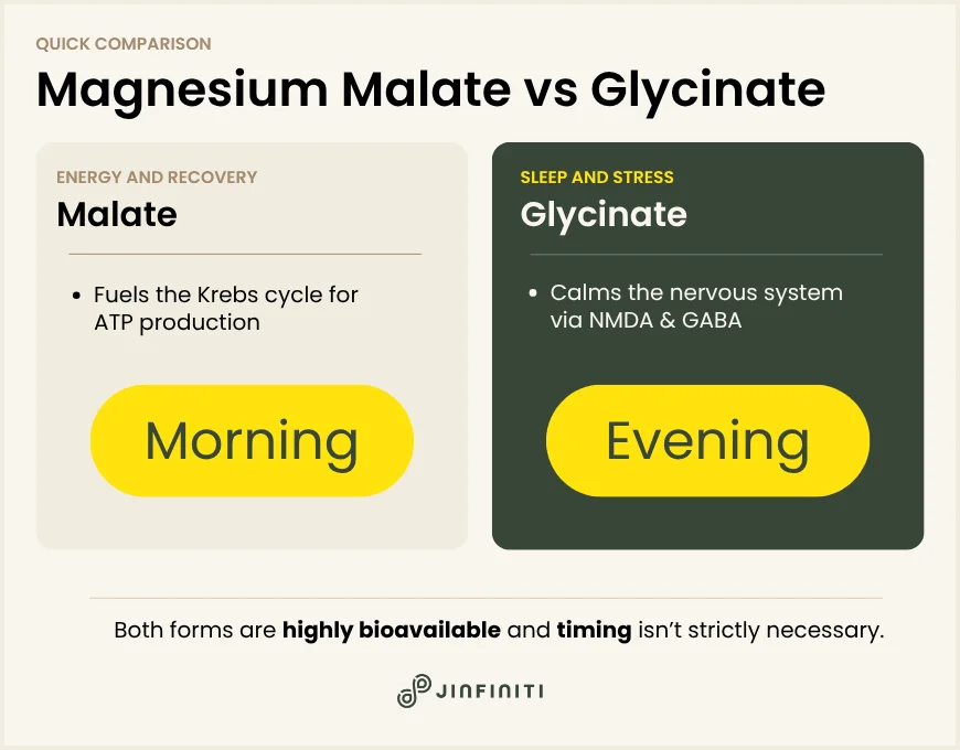
  • Magnesium malate is bonded with malic acid, a compound involved in ATP production; it’s typically better suited for energy and muscle recovery
  • Magnesium bisglycinate is bonded with two glycine molecules that help calm the nervous system; it’s better suited for sleep and stress
  • Both forms are well-absorbed; the meaningful difference is what the bonded compound adds beyond the magnesium itself
  • Using both forms at different times of day is a valid approach, and some supplements are formulated to include both

Magnesium Malate and Glycinate: The Differences

Both malate and glycinate (often appearing on labels as bisglycinate) are organic, chelated forms of magnesium — meaning the mineral is bonded to another compound to improve absorption and reduce digestive side effects. That’s where the similarity ends. What they’re bonded to shapes how they work.

Infographic showing the difference between magnesium malate and glyicnate

Magnesium Malate: The Energy Form

Magnesium malate pairs magnesium with malic acid, a naturally occurring compound found in apples and most fruits. Malic acid is a substrate in the Krebs cycle — the chain of reactions your mitochondria use to generate ATP, the form of energy your cells actually run on. When malate is absorbed alongside magnesium, both compounds become available to support energy production from different angles simultaneously.

That mechanism is part of why magnesium malate has been studied in the context of fibromyalgia, a condition linked to impaired cellular energy and muscle pain. A 1992 study by Abraham and Flechas treated 15 fibromyalgia patients with combined magnesium malate supplementation for 8 weeks and saw meaningful reductions in tender point scores, with some participants reporting improvement within 48 hours. A subsequent double-blind crossover trial by Russell et al. produced more mixed results, showing no significant improvement in the blinded phase. The fibromyalgia evidence isn’t settled.

What is consistent across the research: malate supports ATP-related processes, and people who experience low daytime energy, persistent muscle soreness, or post-exertional fatigue tend to respond better to this form. It’s energizing enough that most practitioners recommend taking it in the morning or early afternoon.

Magnesium Glycinate: The Calm Form

Magnesium glycinate bonds magnesium to two glycine molecules. Glycine is an inhibitory neurotransmitter — it interacts with NMDA receptors in the brain in a way that reduces neural excitability, and research suggests it may lower core body temperature, which is part of what initiates healthy sleep onset.

A 2025 randomized controlled trial in 153 participants with poor sleep quality found that magnesium bisglycinate improved insomnia symptoms compared to placebo, though the effect was modest. A broader systematic review of 15 interventional trials concluded that the majority showed improvement in at least one sleep or anxiety parameter with magnesium supplementation.

The glycine component also appears to have some tissue-specific effects. Animal data suggest bisglycinate may increase brain magnesium concentrations more than other forms — which may explain why it tends to have a more pronounced effect on mood and nervous system symptoms even at comparable doses.

Digestively, bisglycinate is one of the gentlest forms available. It bypasses the osmotic mechanism that makes forms like oxide or citrate laxative at higher doses.

Absorption: Is One Form More Bioavailable?

Magnesium marketing often implies that one form is dramatically more bioavailable than another. The reality is more nuanced.

Both malate and glycinate are chelated organic forms, and research on magnesium bioavailability suggests most organic salts are absorbed at similar rates in the small intestine. Bisglycinate’s edge, if any, appears tissue-specific: it may deliver magnesium to brain tissue more efficiently, whereas its effect on muscle magnesium levels is minimal.

Malate’s advantage isn’t absorption — it’s the malic acid itself. Malic acid contributes to energy metabolism independently of the magnesium it carries. You’re effectively getting two functional compounds in one, which is why malate tends to be the preferred form for people dealing with fatigue or muscle-related symptoms.

For most people, the absorption difference between these two forms isn’t the deciding factor. What matters more is matching the form to the goal.

Magnesium Malate vs. Bisglycinate: Quick Comparison

Magnesium malateMagnesium bisglycinate
Bonded withMalic acidTwo glycine molecules
Primary mechanismKrebs cycle / ATP synthesisNMDA/GABA nervous system support
Best timingMorning or early afternoonEvening
Best forEnergy, muscle fatigue, recoverySleep, stress, anxiety
Digestive toleranceGoodVery gentle
Research baseFibromyalgia, exercise recoverySleep quality, anxiety reduction

Which Magnesium Form Should You Take?

Most comparison guides end here: pick based on your goal. That advice isn’t wrong, but it skips a step.

Around 48% of Americans don’t meet the RDA for magnesium through diet alone. But magnesium deficiency produces symptoms that overlap with a lot of other things — thyroid dysfunction, low NAD+, chronic stress, poor sleep hygiene. Supplementing the right form helps. Supplementing when something else is driving the symptoms doesn’t move the needle.

So: what’s the most likely driver?

⚡Choose Magnesium Malate If You’re Dealing With…

  • Low daytime energy that sleep doesn’t fix
  • Muscle soreness that lingers longer than it should after exercise
  • Fatigue without a clear cause, especially mid-afternoon
  • Fibromyalgia or chronic muscle pain (with the caveat that the evidence here is promising but mixed)

💤Choose Magnesium Glycinate If You’re Dealing With…

  • Trouble falling or staying asleep
  • An overactive mind at night
  • Stress or anxiety that feels like a background hum you can’t quiet
  • Digestive sensitivity to other magnesium forms

Consider Using Both Forms

Taking malate in the morning and bisglycinate in the evening is a strategy some practitioners recommend, and it makes biological sense. You’re supporting energy production during the day and nervous system recovery at night.

This is actually how Jinfiniti’s Vital Minerals Complex is formulated: 240mg of dual-form magnesium combining malate and glycinate in a single supplement. Rather than treating malate and bisglycinate as competing options, the formula treats them as complementary. If you’re not sure which you need — or suspect you need both — that’s the more practical starting point.

For a closer look at how magnesium forms compare across other common pairings, see our guide on magnesium glycinate vs. citrate.

The Magnesium-Energy Connection Most People Miss

Persistent fatigue is one of the most common reasons people turn to magnesium in the first place. But magnesium doesn’t work in isolation.

Magnesium is a cofactor in over 300 enzymatic reactions — including several involved in NAD+ metabolism. NAD+ (nicotinamide adenine dinucleotide) is the molecule your mitochondria depend on to generate ATP that powers cellular function. When magnesium levels are low and NAD+ levels are low at the same time, the energy shortfall comes from two directions. No amount of malate will fully compensate for depleted NAD+.

“Magnesium and NAD+ work hand in hand at the mitochondrial level,” says Dr. Jin-Xiong She, founder of Jinfiniti Precision Medicine. “Addressing one without the other is like fixing one flat tire on a car that has two.”

That’s part of why some people try magnesium, feel a modest improvement, and plateau. Magnesium may be contributing — but it may not be the whole picture. If energy doesn’t recover meaningfully with supplementation, NAD+ deficiency is often the next variable to investigate. It’s also one of the few intracellular deficiencies you can actually test for before supplementing.

For a fuller picture of what supports mitochondrial energy production, see: The Best Mitochondrial Supplements.

Frequently Asked Questions

Is Magnesium Bisglycinate the Same as Magnesium Glycinate?

Yes. The terms are used interchangeably. Both refer to magnesium chelated with two glycine molecules. “Bisglycinate” specifies that there are two glycine molecules bonded to the magnesium (bi = two); “glycinate” is the shorthand most commonly used in supplement marketing. Same compound, different label conventions.

Can I Take Magnesium Malate and Bisglycinate Together?

Yes, and for many people it makes practical sense. Taking malate in the morning and bisglycinate in the evening aligns each form with its primary function — energy support during the day, nervous system recovery at night. Some supplements, including Jinfiniti’s Vital Minerals Complex, combine both forms in a single daily formula.

When Should I Take Magnesium Malate vs. Bisglycinate?

Malate is best taken in the morning or early afternoon. Its energizing properties can interfere with sleep if taken close to bedtime. Bisglycinate is best taken in the evening, where it supports the nervous system wind-down and sleep onset. If you’re using a combined supplement, taking it with dinner is a reasonable middle ground.

Which Form Is Easiest on the Stomach?

Bisglycinate. It bypasses the osmotic laxative effect that makes magnesium oxide and magnesium citrate problematic at higher doses. Malate is also well-tolerated for most people, but bisglycinate is generally the first recommendation for anyone with a sensitive digestive system.

Can Magnesium Supplements Cause Side Effects?

At normal doses (under 350mg of supplemental magnesium per day, per the National Institutes of Health’s tolerable upper intake level), magnesium supplements are well-tolerated for most adults. High doses of oxide and citrate can cause loose stools or cramping. Both malate and bisglycinate are considerably gentler. Anyone with kidney disease should talk with a clinician before supplementing, as impaired kidneys can’t clear excess magnesium efficiently.

  • Abraham, G. E., & Flechas, J. D. (1992). Management of fibromyalgia: Rationale for the use of magnesium and malic acid. Journal of Nutritional Medicine, 3(1), 49–59. https://doi.org/10.3109/13590849208997961
  • Arab, A., Rafie, N., Amani, R., & Shirani, F. (2022). The role of magnesium in sleep health: A systematic review of available literature. Biological Trace Element Research, 201(1), 121–128. https://doi.org/10.1007/s12011-022-03162-1
  • National Institutes of Health, Office of Dietary Supplements. (2022). Magnesium: Fact sheet for health professionals. https://ods.od.nih.gov/factsheets/Magnesium-HealthProfessional/
  • Rawji, A., Peltier, M. R., Mourtzanakis, K., Awan, S., Rana, J., Pothen, N. J., & Afzal, S. (2024). Examining the effects of supplemental magnesium on self-reported anxiety and sleep quality: A systematic review. Cureus, 16(4), e59317. https://doi.org/10.7759/cureus.59317
  • Russell, I. J., Michalek, J. E., Flechas, J. D., & Abraham, G. E. (1995). Treatment of fibromyalgia syndrome with Super Malic: A randomized, double blind, placebo controlled, crossover pilot study. The Journal of Rheumatology, 22(5), 953–958. https://pubmed.ncbi.nlm.nih.gov/8587088/
  • Schuster, J., Cycelskij, I., Lopresti, A., & Hahn, A. (2025). Magnesium bisglycinate supplementation in healthy adults reporting poor sleep: A randomized, placebo-controlled trial. Nature and Science of Sleep, 17, 2027–2040. https://doi.org/10.2147/NSS.S524348
Pomegranate inside a sphere representing Urolithin A vs NAD+

Urolithin A vs NAD+: How They Actually Compare

What You Should Know

  • Urolithin A activates mitophagy, the process that clears out damaged mitochondria; NAD+ fuels the healthy ones that remain
  • They work through separate biological pathways and address different sides of mitochondrial health
  • NAD+ levels can be measured precisely with an at-home intracellular test; urolithin A activity cannot
  • Most people have insufficient NAD+ levels before adding mitophagy support — making NAD+ optimization the logical starting point

Somewhere between the longevity podcasts and the supplement reviews, urolithin A graduated from obscure compound to mainstream recommendation. For people already taking NAD+ precursors, the question followed quickly: are these doing the same thing? Should I add urolithin A? Am I doubling up on something, or covering something new?

They’re covering something new. Urolithin A and NAD+ work through different biological mechanisms, on different timelines, with different evidence bases behind them — and understanding how they diverge changes how you’d sequence either one.

CLIA-Certified NAD Blood Test

Find out if your NAD+ is in the healthy range.

Jinfiniti Intracellular NAD Test on purple background

What Each One Actually Does

Both urolithin A and NAD+ relate to mitochondrial health. That’s where the similarity mostly ends. They operate at completely different stages of the mitochondrial lifecycle, and conflating them leads to poorly sequenced supplementation.

Infographic showing the differences between Urolithin A and NAD+

Urolithin A: The Cleanup Signal

Urolithin A is a postbiotic — not a supplement in the traditional sense, but a compound your gut bacteria produce when they metabolize ellagitannins, polyphenols found in foods like pomegranate, walnuts, and certain berries. The catch is that only about 30–40% of people have the right gut bacteria to convert efficiently. Most people consuming pomegranate juice aren’t producing meaningful levels of urolithin A at all.

Its primary function is activating mitophagy — your cells’ built-in quality control system for mitochondria. Translation: it signals your body to find the damaged, underperforming mitochondria, break them down, and replace them with new ones.

The specific mechanism runs through the PINK1/Parkin pathway: damaged mitochondria are flagged, tagged for removal, cleared out, and recycled. Think of it as the inspection program running in the background of your cells. Old, inefficient units get removed so the network as a whole performs better.

The functional benefits show up slowly. In a 2022 randomized clinical trial published in JAMA Network Open, 66 older adults taking 1,000 mg of urolithin A daily for four months showed significant improvements in both hand and leg muscle endurance compared to placebo, along with reductions in inflammatory and mitochondrial stress biomarkers.

A separate 2022 trial in Cell Reports Medicine found roughly 12% improvements in leg muscle strength in middle-aged adults over a similar timeframe. Cellular gene signatures appear within about four weeks; functional changes tend to emerge at eight to sixteen weeks.

NAD+: The Fuel That Keeps the System Running

NAD+ (nicotinamide adenine dinucleotide) is a coenzyme present in every cell. It shuttles electrons between molecules in the reactions that produce ATP — the energy currency your cells run on. Without sufficient NAD+, mitochondria can’t generate energy efficiently, even if they’re structurally intact.

NAD+ also serves as a substrate for sirtuins and PARPs — enzyme families responsible for DNA repair, stress resistance, and metabolic regulation. Translation: NAD+ isn’t only about energy. It’s also the signal that tells your cells to repair damage, manage inflammation, and maintain function under stress. No NAD+, no repair signal.

The problem is decline. NAD+ levels drop measurably with age, often beginning in the 30s and continuing throughout life. That decline tracks closely with the fatigue, cognitive fog, and slower recovery that many people start noticing in midlife — and that standard bloodwork rarely explains. NMN and NR raise cellular NAD+ levels, typically within days to weeks of starting supplementation.

How They Compare, Side by Side

AspectUrolithin ANAD+
Primary actionClears damaged mitochondria (mitophagy)Fuels energy production and DNA repair
Works throughPINK1/Parkin mitophagy pathwayRedox reactions, sirtuins, PARPs
Speed of effect8–16 weeks for functional changesDays to weeks for NAD+ elevation
Measurable?No consumer test existsYes — intracellular blood spot test
Food sourcesPomegranate, walnuts, berriesNiacin, NMN, NR precursors
Gut dependencyYes (conversion varies widely)No
Best evidence forMuscle endurance, mitochondrial qualityCellular energy, cognitive function, repair

One of These Can Be Measured. The Other Can’t.

You can measure one of these. You can’t measure the other.

Intracellular NAD+ levels can be tested directly — from a finger-prick blood spot sample, processed through a CLIA-certified lab — and the results tell you exactly where you stand. You can test before starting supplementation, again at four to eight weeks, and know with precision whether your intervention is working and whether your dose needs adjusting.

Urolithin A has no equivalent. There’s no at-home test that tells you whether your mitophagy is active, how efficiently your gut is converting, or whether the supplement you’re taking is doing anything measurable. The clinical evidence for urolithin A is real and encouraging, but the individual feedback loop simply doesn’t exist yet.

Supplementation without measurement is guesswork. You can spend months on a stack you believe is working with no objective signal either way.

“Most people come to us having already tried several supplements, often including NAD+ precursors, without ever testing their baseline,” says Dr. Jin-Xiong She, founder of Jinfiniti Precision Medicine and a genomic scientist with over 400 peer-reviewed publications. “When we test them, a significant portion are still deficient. The supplement was real — but the dose or formulation wasn’t moving their levels into the optimal range. That’s what testing reveals.”

Can They Work Together?

They can, and the case for combining them is straightforward: they address two different phases of the mitochondrial lifecycle. Urolithin A removes underperforming mitochondria; NAD+ fuels and maintains the ones that remain. One clears, the other powers. Those aren’t redundant functions — they’re sequential ones.

Some preclinical work suggests urolithin A may also modestly elevate NAD+ levels by improving overall mitochondrial efficiency — but this has only been observed in animal models so far. Human data confirming that effect isn’t available yet.

What the Research Supports (and What It Doesn’t)

There are no head-to-head human trials directly comparing urolithin A and NAD+ supplementation. The strongest clinical evidence for urolithin A is in muscle endurance and mitochondrial biomarkers in older and middle-aged adults. For NAD+ precursors, Jinfiniti’s clinical data showed 85% of participants reaching optimal intracellular NAD+ levels within four weeks of using the Vitality↑® NAD+ Booster — with average levels doubling — which is among the most rigorous published results in consumer longevity supplementation.

A 2024 study in Frontiers in Aging Neuroscience compared urolithin A and nicotinamide riboside (NR) in human microglial cells and found that both reduced DNA damage-induced cellular senescence, though through different pathways — a finding that supports the case for combining them rather than treating them as interchangeable.

How to Think About Sequencing

If your NAD+ levels are deficient — which is common and often invisible without testing — adding urolithin A on top of a fuel-depleted system is like cleaning a factory floor before turning the power back on. The cleanup matters, but it can’t compensate for missing energy.

Establishing an optimal NAD+ baseline first gives your mitochondria the fuel they need to function well. Once that’s verified and stable, urolithin A becomes a logical complement — one that improves the quality of the mitochondrial network your NAD+ is now powering effectively.

Which One Should You Prioritize?

If you don’t know your NAD+ baseline, start there. NAD+ deficiency is common, measurable, and often the root cause of energy and cognitive complaints that other bloodwork misses. Testing first tells you whether your levels are deficient, suboptimal, or already in range — and that answer changes what you do next.

If your NAD+ levels are already confirmed optimal and you’re focused on muscle endurance, exercise recovery, or longer-range mitochondrial quality as you age, urolithin A is a well-supported complement.

For most people doing this seriously, the goal is eventually both. The order matters more than people assume — and measurement is what makes the sequence rational instead of guessed at.

Frequently Asked Questions

Is urolithin A the same as NAD+?

No. They’re chemically distinct and work through separate pathways. Urolithin A is a gut-derived postbiotic that activates mitophagy — the removal of damaged mitochondria. NAD+ is a coenzyme that fuels energy production and activates DNA repair enzymes. They address complementary but different aspects of cellular health.

Does urolithin A raise NAD+ levels?

Animal studies suggest it may modestly, by improving mitochondrial efficiency — but this hasn’t been confirmed in human trials. It’s not a substitute for direct NAD+ supplementation if your levels are deficient.

Can I take urolithin A and NMN together?

Yes. They target different parts of the mitochondrial lifecycle and are generally considered complementary. NMN supports energy production and sirtuin activity; urolithin A supports mitochondrial quality control. No known interaction between them.

How long does urolithin A take to work compared to NAD+?

NAD+ precursors can elevate cellular NAD+ within days to a few weeks. Urolithin A works on a slower cadence — cellular gene signatures appear around four weeks, functional changes in endurance and strength typically at eight to sixteen weeks, based on the 2022 clinical trials.

How do I know if my NAD+ levels are optimal?

An intracellular NAD+ test measures your actual cellular NAD+ concentration from a finger-prick blood sample and tells you whether you’re in the optimal range (40–100 μM), suboptimal, or deficient. It’s the only objective way to know whether your supplementation is working — and how much to adjust.

  • Liu, S., D’Amico, D., Shankland, E., Bhayana, S., Garcia, J. M., Aebischer, P., Rinsch, C., Singh, A., & Marcinek, D. J. (2022). Effect of urolithin A supplementation on muscle endurance and mitochondrial health in older adults: A randomized clinical trial. JAMA Network Open, 5(1), e2144279. https://doi.org/10.1001/jamanetworkopen.2021.44279
  • Ryu, D., Mouchiroud, L., Andreux, P. A., Katsyuba, E., Moullan, N., Nicolet-dit-Félix, A. A., Williams, E. G., Jha, P., Lo Sasso, G., Huzard, D., Aebischer, P., Sandi, C., Rinsch, C., & Auwerx, J. (2016). Urolithin A induces mitophagy and prolongs lifespan in C. elegans and increases muscle function in rodents. Nature Medicine, 22(8), 879–888. https://doi.org/10.1038/nm.4132
  • Singh, A., D’Amico, D., Andreux, P. A., Dunngalvin, G., Kern, T., Blanco-Bose, W., Aebischer, P., Auwerx, J., & Rinsch, C. (2022). Urolithin A improves muscle strength, exercise performance, and biomarkers of mitochondrial health in a randomized trial in middle-aged adults. Cell Reports Medicine, 3(5), 100633. https://doi.org/10.1016/j.xcrm.2022.100633
  • Serantes, D., Muñoz-Guardiola, P., Megías-Roda, E., García-Martinez, I., Morales, M., Santamaria-Martínez, A., Segués, N., Rossignol, J., Galobart, R., Romanos, G., Esteve-Codina, A., Vidal-Alabró, A., Ingham, E., Muñoz, J., & Mulero, M. (2024). Urolithin A and nicotinamide riboside differentially regulate innate immune defenses and metabolism in human microglial cells. Frontiers in Aging Neuroscience, 16, 1462752. https://doi.org/10.3389/fnagi.2024.1462752
Minimalist illustration of a translucent battery shape filled with glowing cellular mitochondria representing NAD

What Is NAD and Why Does It Matter for Your Energy?

What You Should Know

  • NAD is a coenzyme that turns food into cellular energy and supports DNA repair
  • Your levels naturally decline with age—often beginning in your 30s
  • Low NAD contributes to fatigue, brain fog, and slower recovery
  • Without testing, you can’t know if your levels are deficient or if supplementation is working

When exhaustion doesn’t respond to the standard fixes, it often points to something happening at the cellular level. One of the most common and underrecognized drivers is a decline in NAD, a molecule your cells rely on to produce energy.

NAD — short for nicotinamide adenine dinucleotide — exists in every living cell in your body. It’s not a vitamin or a hormone. It’s a coenzyme, which means it helps other molecules do their jobs. Without sufficient NAD, your cells can’t convert the food you eat into usable energy. They also can’t repair DNA damage, respond to stress, or regulate metabolism effectively.

The problem is that NAD levels decline measurably with age, often beginning in your 30s or 40s. Research shows that many people experience a 40-50% drop by age 50. And unlike most biomarkers, NAD can be measured directly — which means you can know exactly where your levels stand and take action based on data, not guesswork.[1]

CLIA-Certified NAD Blood Test

Find out if your NAD+ is in the healthy range.

Jinfiniti Intracellular NAD Test on purple background

What NAD Actually Is

NAD is a small molecule found in every living cell, from bacteria to humans. It’s been studied for over a century — first discovered in 1906 during fermentation research — but our understanding of its importance in human health has deepened significantly in recent decades.

At a chemical level, NAD is made of two nucleotides joined by phosphate groups. One contains adenine, the other nicotinamide. The body can produce NAD from vitamin B3 (niacin) or the amino acid tryptophan, though most NAD is recycled through a salvage pathway rather than built from scratch.

What makes NAD essential is its ability to shuttle electrons between molecules. This electron transfer is the basis for nearly all energy production in your body.

The Two Forms: NAD+ and NADH

NAD exists in two forms that constantly cycle back and forth: NAD+ and NADH.

NAD+ is the oxidized form — think of it as an empty shuttle bus, ready to pick up electrons during metabolic reactions. NADH is the reduced form — the shuttle carrying electrons that can be used to generate ATP, your cells’ energy currency.

Together, these forms create a loop. NAD+ accepts electrons from nutrients like glucose and fat, becoming NADH. NADH then delivers those electrons to the mitochondria, where they drive ATP production. Once the electrons are dropped off, NADH converts back to NAD+, and the cycle continues.

This constant cycling is what keeps energy flowing in your cells. When NAD levels are too low, the loop slows down. Cells struggle to produce enough ATP, and you feel it as fatigue, slower recovery, or cognitive fog.

Why NAD Matters: What It Does in Your Body

NAD is involved in hundreds of cellular processes. Studies show it participates in more than 500 enzymatic reactions.[2]

A few roles stand out as particularly critical:

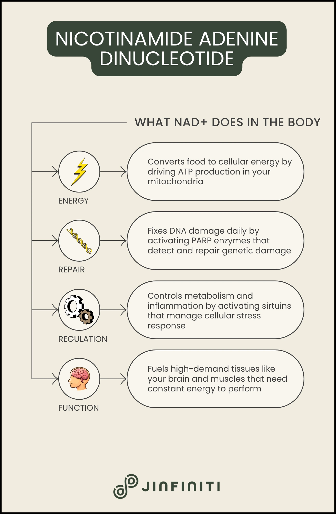
Energy production: Inside your mitochondria, NAD+ drives the reactions that convert glucose and fatty acids into ATP. Without sufficient NAD, mitochondrial function declines, and cellular energy production becomes less efficient.

DNA repair: Every day, your cells accumulate DNA damage from normal metabolism, UV exposure, and environmental toxins. NAD is consumed by enzymes called PARPs that detect and repair this damage. When NAD levels are low, DNA repair slows, and damage accumulates faster.

Metabolic regulation: A family of proteins called sirtuins only function in the presence of NAD. Sirtuins influence everything from fat storage to inflammation to circadian rhythm. They’re often called “longevity genes” because of their connection to healthy aging and stress resistance.

Brain function: Your brain represents only about 2% of your body weight but consumes roughly 20% of your total energy. NAD supports the high metabolic demands of neurons, and declining NAD in the brain has been linked to cognitive decline and neurodegenerative conditions.[3]

This isn’t just about “anti-aging.” NAD is how your body functions today. When levels are optimal, cells repair themselves efficiently, produce energy consistently, and respond to stress effectively. When levels drop, those processes slow — and you feel the difference before any disease state appears.

Infographic showing what NAD (Nicotinamide Adenine Dinucleotide) does in the body

Why NAD Levels Decline With Age

NAD decline is one of the most consistent findings in aging research. It’s been documented in rodents, primates, and humans across multiple tissues.

Human studies show that whole blood NAD levels decline significantly with age, particularly in men. Women’s levels tend to fluctuate more after age 50, likely due to hormonal changes. In skeletal muscle, the decline can be anywhere from 15% to 65%, depending on the individual and the measurement method. Brain NAD also decreases measurably, which correlates with reduced cognitive function.[1]

The decline happens through two main mechanisms.

Reduced production occurs because the enzymes responsible for making NAD become less efficient with age. The key enzyme, NAMPT, decreases in activity over time. This creates a bottleneck — your cells can’t produce NAD as quickly as they once did, even if you’re consuming adequate precursors through diet.[4]

Increased consumption happens because certain NAD-degrading enzymes become more active as you age. One called CD38 rises in response to chronic inflammation. Another group, PARPs, ramps up activity in response to accumulated DNA damage. Both consume NAD to do their jobs, which depletes the cellular pool.[5][6]

Think of it as a sink where the faucet slows while the drain widens. Less NAD coming in, more NAD going out. Over time, this leads to the net decline that begins earlier than most people realize.

What Drives NAD Decline?

Age is the primary driver, but several factors accelerate the process:

  • Chronic inflammation increases the activity of CD38 and other NAD-consuming enzymes[5]
  • Metabolic stress from a high-fat or high-sugar diet can reduce skeletal muscle NAD levels in just two months[7]
  • Obesity is associated with both lower NAMPT expression and higher PARP activity, creating a compounding effect[8]
  • DNA damage from UV exposure, toxins, or normal cellular metabolism forces PARPs to consume more NAD for repair[6]

If you’re dealing with persistent fatigue despite normal blood tests, NAD deficiency is one of the cellular-level explanations that standard panels miss.

Signs Your NAD Levels May Be Low

NAD decline doesn’t announce itself with a single, obvious symptom. Instead, it shows up as a pattern of issues that conventional testing often fails to explain.

Common signs of low NAD include:

  • Persistent fatigue that doesn’t resolve after adequate sleep — not the kind that goes away after a good night’s rest, but the kind that lingers despite doing everything “right”
  • Slower recovery from exercise, illness, or stress — when NAD levels are optimal, cells repair damage efficiently and bounce back quickly; when levels are low, recovery takes longer
  • Brain fog and difficulty concentrating — neurons are metabolically demanding, and when NAD drops, cognitive performance often follows
  • Metabolic changes — difficulty maintaining weight, worsening insulin sensitivity, or changes in how your body responds to food

If you’re still tired after 8 hours of sleep, cellular energy deficiency is worth investigating.

The challenge is that these symptoms overlap with many other conditions. Thyroid disorders, sleep apnea, anemia, hormonal imbalances, and dozens of other issues can present similarly. That’s why symptom-based guessing rarely leads to a clear answer.

Measurement is the only way to know.

The NAD Measurement Problem

Most people who take NAD supplements have no idea if they’re actually working.

The wellness industry has embraced NAD precursors like NMN and NR over the past few years. Hundreds of products now claim to boost NAD levels, often with impressive-sounding promises about energy, longevity, and cognitive performance. But very few people taking these supplements ever test their levels — before, during, or after.

That’s a problem, because NAD metabolism is highly individual. The same dose that optimizes one person’s levels might do almost nothing for another. Some people absorb precursors efficiently; others don’t. Some have high baseline consumption from inflammation or DNA damage; others don’t. Without testing, you’re guessing.

Intracellular measurement matters because that’s where NAD actually works. Some tests measure NAD in blood plasma, which can give you a general sense of systemic levels. But the NAD inside your cells — where energy production, DNA repair, and metabolic regulation happen — is what drives outcomes. Intracellular NAD testing provides a much more accurate picture of your cellular energy status.

Dr. Jin-Xiong She’s clinical research established clear benchmarks for interpreting results. Optimal NAD levels fall between 40 and 100 micromolar (μM). Anything below 40μM is considered suboptimal or deficient. Levels above 100μM may not provide additional benefits and could potentially cause issues.

These ranges give you a concrete target, not a vague reference to “normal.”

Why Testing Changes Everything

Jinfiniti developed the world’s first consumer-grade intracellular NAD test in 2019. Before that, NAD testing was largely confined to research labs. Now, you can measure your baseline, track changes over time, and personalize your approach based on data.

Here’s what measurement enables:

  • Establishing your baseline tells you where you’re starting from — you might discover your levels are already optimal, in which case supplementation isn’t needed, or you might find they’re severely deficient, which explains symptoms that have gone undiagnosed for years
  • Validating whether supplementation works removes the guesswork — some people respond well to NMN, others respond better to niacinamide or NR, and testing shows you what’s actually happening in your cells, not what a bottle label promises
  • Personalizing your dosing is critical because there’s no one-size-fits-all approach to NAD optimization — in Jinfiniti’s clinical trial, 85% of participants reached optimal NAD levels within four weeks, but that success rate depended on personalized dosing based on measured results, not generic recommendations
  • Tracking progress over time lets you adjust as needed — NAD levels aren’t static; they respond to changes in diet, exercise, stress, inflammation, and supplementation, so retesting every few months ensures you’re maintaining optimal levels rather than drifting back into deficiency

As Dr. She puts it: “Measure before acting. NAD optimization isn’t about taking the same dose as someone else and hoping it works. It’s about knowing your levels, acting on that information, and confirming the results. Precision requires data.”

Without measurement, you’re flying blind. With it, you can make informed decisions and track real progress.

How to Support Healthy NAD Levels

If you’re looking to optimize your NAD levels, the most effective approach combines testing with targeted intervention.

Here’s what works:

  • Test first — Establish your baseline with an intracellular NAD test; this removes the guesswork and tells you whether your levels are deficient, suboptimal, or already optimal
  • NAD+ precursor supplementation is the most direct way to raise levels — NMN and NR are the most studied precursors; both convert to NAD inside your cells, though they follow slightly different pathways; niacinamide (a form of vitamin B3) also works through the salvage pathway
  • Lifestyle factors can support NAD production — regular exercise, moderate caloric intake, and consistent sleep all influence NAD metabolism, though they’re unlikely to fully counter age-related decline on their own
  • Reduce chronic inflammation through diet, stress management, and addressing underlying health issues — this helps by lowering NAD consumption from enzymes like CD38
  • Retest and adjust after 4-6 weeks — if your levels haven’t reached the optimal range, adjust your dose or try a different precursor; if they have, continue monitoring periodically to ensure they stay stable

Jinfiniti’s Vitality NAD+ Booster uses a multi-pathway formula that combines NMN, niacinamide, creatine, and D-ribose. The clinical trial data showed an average doubling of NAD levels within four weeks, with 85% of participants reaching the optimal range.

The goal isn’t to chase an arbitrary number. It’s to restore your cells’ ability to produce energy efficiently, repair damage effectively, and respond to stress appropriately. When NAD levels are optimal, you feel it — and the data confirms it.

The Bottom Line

NAD is not a wellness trend. It’s a fundamental molecule that’s been studied for over a century and plays a documented role in energy production, DNA repair, and metabolic regulation. Levels decline measurably with age, and that decline has real consequences — fatigue, slower recovery, cognitive fog, and increased vulnerability to age-related disease.

The difference between vague health advice and precision medicine comes down to measurement. You can’t optimize what you don’t measure. And without data, you’re guessing whether your NAD levels are deficient, whether supplementation is working, or whether you even need it in the first place.

If you’re dealing with unexplained fatigue, cognitive decline, or signs of accelerated biological aging, NAD testing gives you a clear starting point. It’s not the only factor that matters, but it’s one you can measure, address, and track over time.

Frequently Asked Questions

Is NAD the same as vitamin B3?

NAD is a molecule your body makes from vitamin B3 (which includes niacin, niacinamide, and nicotinamide riboside). Vitamin B3 is a precursor — a building block your cells use to produce NAD. You can get vitamin B3 from food or supplements, but your body still needs to convert it into NAD through metabolic pathways. Learn more about the differences between NAD, niacin, and niacinamide.

Which foods contain NAD?

Very few foods contain NAD directly. Instead, you get NAD precursors from foods rich in vitamin B3 — like meat, fish, mushrooms, peanuts, and fortified grains — or from foods containing tryptophan, an amino acid your body can convert to NAD. However, dietary sources alone rarely raise NAD levels significantly, especially after age 40. See the full list of NAD-boosting foods.

Are NAD supplements safe?

NAD precursors like NMN, NR, and niacinamide are generally well-tolerated in clinical studies, with mild side effects like nausea or flushing reported in some people. Most research shows a favorable safety profile for short-term use. However, long-term safety data is still limited, and it’s best to work with a healthcare provider — especially if you have underlying health conditions.

Can NAD help with weight loss?

NAD doesn’t directly cause weight loss, but it plays a role in metabolic function and energy regulation. Some research suggests NAD precursors may support metabolic health, particularly when combined with exercise and caloric moderation. However, NAD supplementation is not a weight-loss drug, and results vary widely between individuals. Read more about NAD, NMN, and weight loss.

What is the difference between NAD+ and NADH?

NAD+ is the oxidized form of the molecule, ready to accept electrons during metabolic reactions. NADH is the reduced form, carrying electrons that can be used to generate ATP. They cycle back and forth continuously, and both are necessary for cellular energy production.

Can you increase NAD levels naturally without supplements?

Lifestyle factors like exercise, caloric moderation, and adequate sleep can support NAD production to some degree. However, research suggests that lifestyle changes alone are often insufficient to fully counter the age-related decline in NAD levels, particularly after age 40. Precursor supplementation is typically needed for meaningful increases.[2]

What are optimal NAD levels?

Based on Dr. Jin-Xiong She’s clinical research, optimal intracellular NAD levels fall between 40 and 100 micromolar (μM). Levels below 40μM are considered suboptimal or deficient. Levels above 100μM may not provide additional benefits and could potentially be harmful.

How do I know if my NAD levels are low?

The only reliable way to know is through testing. Symptoms like persistent fatigue, brain fog, and slow recovery can suggest low NAD, but they overlap with many other conditions. Intracellular NAD testing provides a precise measurement of your cellular levels.

Does NAD supplementation work for everyone?

NAD precursors can raise levels in most people, but individual responses vary widely. Some people absorb and convert precursors efficiently, while others need higher doses or different forms. Without testing, you won’t know if a supplement is actually working for you. That’s why personalized dosing based on measured levels is more effective than generic recommendations.

How long does it take to see results from NAD supplementation?

Most people who respond to NAD precursors notice changes within 2-4 weeks. Clinical studies, including Jinfiniti’s trial with Vitality NAD+ Booster, show measurable increases in NAD levels within four weeks. However, subjective improvements in energy, recovery, and cognitive clarity may appear sooner or later depending on your baseline levels and overall health.

Does NAD interact with GLP-1 medications?

NAD and GLP-1 pathways both influence metabolic health, and there’s emerging research on how they interact. If you’re using GLP-1 medications, NAD optimization may offer complementary benefits. Learn more about the relationship between NAD and GLP-1.

  1. Yang F, Deng X, Yu Y, Luo L, Chen X, Zheng J, et al. Association of Human Whole Blood NAD+ Contents With Aging. Frontiers Media SA; 2022. https://doi.org/10.3389/fendo.2022.829658
  2. Covarrubias AJ, Perrone R, Grozio A, Verdin E. NAD+ metabolism and its roles in cellular processes during ageing. Springer Science and Business Media LLC; 2020. https://doi.org/10.1038/s41580-020-00313-x
  3. Zhu XH, Lu M, Lee BY, Ugurbil K, Chen W. In vivo NAD assay reveals the intracellular NAD contents and redox state in healthy human brain and their age dependences. Proceedings of the National Academy of Sciences; 2015. https://doi.org/10.1073/pnas.1417921112
  4. Zhang W, Ren H, Chen W, Hu B, Feng C, Li P, et al. Nicotinamide phosphoribosyltransferase in NAD+ metabolism: physiological and pathophysiological implications. Springer Science and Business Media LLC; 2025. https://doi.org/10.1038/s41420-025-02672-w
  5. Piedra-Quintero ZL, Wilson Z, Nava P, Guerau-de-Arellano M. CD38: An Immunomodulatory Molecule in Inflammation and Autoimmunity. Frontiers Media SA; 2020. https://doi.org/10.3389/fimmu.2020.597959
  6. Sousa FG, Matuo R, Soares DG, Escargueil AE, Henriques JAP, Larsen AK, et al. PARPs and the DNA damage response. Oxford University Press (OUP); 2012. https://doi.org/10.1093/carcin/bgs132
  7. Rasool S, Geetha T, Broderick TL, Babu JR. High Fat With High Sucrose Diet Leads to Obesity and Induces Myodegeneration. Frontiers Media SA; 2018. https://doi.org/10.3389/fphys.2018.01054
  8. Rappou E, Jukarainen S, Rinnankoski-Tuikka R, Kaye S, Heinonen S, Hakkarainen A, et al. Weight Loss Is Associated With Increased NAD+/SIRT1 Expression But Reduced PARP Activity in White Adipose Tissue. The Endocrine Society; 2016. https://doi.org/10.1210/jc.2015-3054
Image of GLP-1 medication next to body tape measure

NAD and GLP-1: What the Research Actually Shows

What You Should Know

  • GLP-1 medications reduce caloric intake and weight, but up to 45% of the weight lost may come from lean body mass rather than fat alone.
  • NAD+ — a molecule found in every cell — fuels the mitochondria that produce cellular energy, and levels typically decline with age and metabolic stress.
  • Animal research suggests that intestinal NAD+ biosynthesis is required for the gut to produce GLP-1 naturally, though human data is still emerging.
  • If you’re supplementing NAD+ alongside a GLP-1 protocol, testing your intracellular NAD+ levels is the only way to know whether supplementation is actually working.

If you’re on a GLP-1 medication — Ozempic, Wegovy, Mounjaro — and you’ve been told to add NAD+ to your protocol, you’ve probably encountered some version of the same pitch: NAD+ gives you the energy back that GLP-1 drugs take away. It’s simple, it sounds plausible, and it’s only partially right.

The real relationship between NAD+ and GLP-1 is more interesting than that. There’s research — not widely discussed in consumer health circles — suggesting that NAD+ doesn’t just patch the side effects of GLP-1 medications. It plays a role in whether your body produces GLP-1 naturally in the first place. That reframes the conversation considerably.

Here’s what the evidence actually supports, where it’s still emerging, and what to consider if you’re thinking about pairing the two.

CLIA-Certified NAD Test Kit

Find out if your NAD+ is actually optimized.

Jinfiniti Intracellular NAD Test on purple background

Why GLP-1 Medications Leave Some People Feeling Depleted

GLP-1 receptor agonists work by mimicking a hormone your intestines naturally produce after eating. They slow gastric emptying, reduce appetite, and signal the brain that you’re full. The result — significant caloric restriction — is exactly what drives the weight loss.

But the body doesn’t distinguish between a medication-induced caloric deficit and a famine. When fuel intake drops, cellular energy production follows.

What Caloric Restriction Does to Cellular Energy

Your cells produce energy through the mitochondria, using a process that requires a steady supply of fuel and specific molecular cofactors — including NAD+. When you eat significantly less, the inputs into that energy production chain decrease. For many people on GLP-1 medications, the experience is fatigue, mental fog, and a general feeling of physical flatness that doesn’t match the number on the scale.

This isn’t a sign the medication isn’t working. It’s a sign the body is adapting to reduced intake — and that adaptation has cellular costs.

The Lean Mass Question

The more pressing clinical concern is what’s happening to body composition. In the STEP-1 clinical trial of semaglutide, roughly 45% of total weight lost was lean body mass rather than fat. A 2025 Cell Metabolism analysis noted that lean mass loss on GLP-1 receptor agonists has raised genuine questions about the long-term metabolic implications — particularly for people already experiencing age-related muscle decline.[1]

Losing muscle alongside fat matters because muscle is metabolically active tissue. Less muscle means a lower resting metabolic rate, reduced physical capacity, and — critically — less mitochondrial machinery to support energy production.

This is the fuller picture of why some people feel depleted on GLP-1 medications. It’s not just caloric restriction. It’s what caloric restriction, at scale and over time, does to the cellular infrastructure that produces energy in the first place.

If you’ve been experiencing that and wondering whether something deeper is going on, you might want to read more about why standard bloodwork often misses this kind of fatigue.

What NAD+ Does — and Why It Matters Here

NAD+ — nicotinamide adenine dinucleotide — is a coenzyme found in every living cell. It’s the molecule that allows your mitochondria to convert food into usable energy, supports DNA repair, and activates a class of proteins called sirtuins that regulate cellular aging and metabolism.

NAD+ levels decline with age — measurably so, beginning in the 30s and accelerating through midlife. They also decline with metabolic stress, obesity, and high-fat diets. This matters for people on GLP-1 medications because the population most likely using these drugs — adults with metabolic dysfunction, insulin resistance, or obesity — often starts from a lower NAD+ baseline to begin with.

When you add significant caloric restriction to an already-depleted NAD+ system, the cellular energy shortfall compounds. This is the specific gap NAD+ supplementation is meant to address.

A 2025 study published in Obesity found that semaglutide-induced weight loss improved mitochondrial oxidative phosphorylation efficiency in skeletal muscle in mice — meaning GLP-1 medications may independently support some mitochondrial improvements through weight loss itself.[2]

That’s genuinely encouraging. But mitochondrial efficiency and NAD+ availability are distinct variables, and the evidence for NAD+ specifically addresses the fuel supply side of the equation.

NAD+ and Your Body’s Own GLP-1 Production

Most articles frame NAD+ as a complement to GLP-1 medications — something you take alongside Ozempic to feel better. The deeper story, supported by emerging research, is that NAD+ may be upstream of GLP-1 production entirely.

How Intestinal NAD+ Controls Natural GLP-1 Production

GLP-1 is produced by specialized L-cells in the lining of your small intestine. Those cells depend on a tightly regulated internal environment to function properly — and a key part of that environment is NAD+ biosynthesis, mediated by an enzyme called NAMPT (nicotinamide phosphoribosyltransferase).

A 2022 study published in Endocrinology by Nagahisa et al. tested what happens when intestinal epithelial cells are stripped of NAMPT — and therefore unable to produce NAD+. The result was a significant reduction in GLP-1 production, decreased insulin secretion, and elevated postprandial blood glucose. The mice weren’t heavier. They just couldn’t regulate glucose properly because their guts couldn’t produce enough GLP-1.[3]

What Happens When Gut NAD+ Is Depleted

The same research team found that obese mice fed a high-fat diet showed the same pattern: compromised intestinal NAD+ biosynthesis, impaired GLP-1 production, and disrupted postprandial glucose metabolism. When they administered NMN — a key NAD+ precursor — intestinal NAD+ levels were restored, and GLP-1 production recovered along with glucose regulation.

A follow-up review in Nutrients (2023) elaborated on the mechanistic pathway: intestinal NAD+ biosynthesis, via the AMPK–NAMPT–SIRT1 axis, is critical for maintaining the gut environment that produces GLP-1. When that pathway is disrupted — by obesity, aging, or poor diet — GLP-1 production downstream suffers.[4]

It’s worth being clear: this research is primarily in animal models. Human clinical trials on this specific pathway are still underway. But the mechanistic insight changes the framing. The metabolic dysfunction that leads people to need GLP-1 medications may be partly downstream of the same NAD+ depletion those medications are compensating for. If that’s accurate, supporting NAD+ isn’t just managing side effects. It’s addressing the cellular environment the whole system depends on.

NAD+ and Muscle Preservation During GLP-1 Treatment

The lean mass concern with GLP-1 medications is real enough to take seriously, and NAD+ has a specific role to play here.

Muscle tissue contains a high density of mitochondria — more than almost any other tissue in the body. Mitochondrial function in muscle depends heavily on NAD+ availability. When NAD+ is depleted, mitochondrial output falls, and muscle cells become less efficient at producing energy and maintaining their own structure.

There’s also a creatine connection worth noting. NAD+ and creatine operate through overlapping metabolic pathways — both supporting ATP synthesis, the actual energy currency inside cells. For people concerned about muscle quality and energy output during GLP-1-induced weight loss, addressing both pathways simultaneously is a reasonable approach. If you want to understand how supplementing each of these nutrients compares in terms of NAD+ dosing specifically, the NAD+ dosage guide covers that in detail.

The 2026 Cell Reports Medicine analysis found that GLP-1 receptor agonist treatment did upregulate certain mitochondrial proteins — including SIRT5 — in skeletal muscle compared to calorie restriction alone. That’s a meaningful finding. But it also underscores that the mitochondrial effects of GLP-1 treatment are still being characterized, and that NAD+ addresses the fuel side of mitochondrial function in a way that GLP-1 doesn’t.

Measuring NAD+ While on GLP-1 Medications

The population on GLP-1 medications tends to be engaged with their health — tracking metrics, working with clinicians, paying attention to how interventions affect outcomes. Adding NAD+ supplementation to a GLP-1 protocol without measuring baseline NAD+ levels is, by the standards of that same engagement, a gap.

Intracellular NAD+ can be measured directly through a finger-prick blood test. Optimal levels, based on Jinfiniti’s clinical research, fall between 40 and 100 μM. Most adults in midlife — particularly those with obesity or metabolic dysfunction — test significantly below that range before supplementing.

“The people most likely to benefit from NAD+ support are often the ones who started below optimal levels without knowing it,” says Dr. Jin-Xiong She, founder of Jinfiniti Precision Medicine. “Supplementing without a baseline is still just guessing — and when you’re already managing a prescription protocol, precision matters more, not less.”

Knowing your starting level also allows you to calibrate dosing appropriately and confirm, after 4–6 weeks of supplementation, whether you’ve actually reached the range where NAD+ exerts its measurable effects. Niacinamide — one of the four ingredients in the Vitality NAD+ Booster — plays a specific role in that synthesis pathway; understanding what niacinamide does in the context of NAD+ production is useful for anyone building a targeted protocol.

What to Realistically Expect From NAD+ Alongside GLP-1 Therapy

NAD+ is not a GLP-1 amplifier. It won’t increase the weight-loss effect of semaglutide or make the medication work faster. What it does is support the cellular environment that gets taxed when caloric intake drops significantly and the body is remodeling its composition.

For people on GLP-1 medications, the realistic case for NAD+ includes: better mitochondrial support during caloric restriction, reduced cellular energy depletion in muscle tissue, and — based on emerging animal research — potential support for the gut environment that produces GLP-1 naturally. None of this is a guarantee. Individual response to NAD+ supplementation varies, and starting NAD+ levels, age, diet, and baseline metabolic health all affect outcomes.

The most important caveat: if you’re on a GLP-1 medication and considering adding NAD+ supplementation, talk with your clinician first. NAD+ precursors are generally well-tolerated, but any addition to an active prescription protocol warrants a conversation with the prescribing provider.

The supplementation question is secondary to the measurement question. Know your NAD+ level. Then decide what to do with that information.

Frequently Asked Questions

Can you take NAD+ with Ozempic, Wegovy, or Mounjaro?

Yes. NAD+ precursors — including NMN and NR — are generally well-tolerated and are not known to interact with GLP-1 receptor agonists. That said, if you’re on a prescription protocol, the appropriate step is to confirm with your prescribing clinician before adding any supplement.

Can you get NAD+ injections while on GLP-1 medications?

Yes. NAD+ injections aren’t known to interact with GLP-1 receptor agonists, and some clinics offer them specifically for GLP-1-related fatigue. One caveat: the NAD+ molecule is too large to enter cells directly from the bloodstream, so IV delivery loses a significant portion to breakdown before it’s usable. Subcutaneous injections perform somewhat better — but Jinfiniti’s clinical data showed their oral multi-pathway formula reached optimal intracellular levels more consistently than subcutaneous NAD+, at a fraction of the cost.

Why do GLP-1 medications cause fatigue, and can NAD+ help?

GLP-1 medications reduce caloric intake substantially, which decreases the fuel available for cellular energy production. NAD+ supports the mitochondrial machinery that converts available fuel into usable energy. For people whose NAD+ levels are already depleted — common in those with metabolic dysfunction — supplementing to optimal levels may help address that energy gap. Results vary based on where you’re starting from.

Does NAD+ affect how GLP-1 medications work?

Not directly. NAD+ and GLP-1 receptor agonists work through different mechanisms. What’s interesting is the upstream relationship: animal research suggests that NAD+ biosynthesis in the gut is required for the body to produce GLP-1 naturally. This doesn’t mean NAD+ boosts the medication — but it does suggest the two systems are more connected at a biological level than the standard framing implies.

Is lean mass loss on GLP-1 medications inevitable?

Data from the STEP-1 trial showed significant lean mass reduction alongside fat loss on semaglutide. Whether this is inevitable or modifiable depends on factors including exercise, protein intake, and mitochondrial support. NAD+ supports muscle mitochondrial function and operates through pathways that overlap with creatine — both relevant to maintaining muscle quality during weight loss.

How do you know if your NAD+ levels are actually low?

You test. Intracellular NAD+ levels can be measured through a finger-prick blood test processed in a CLIA-certified lab. Optimal levels fall between 40 and 100 μM; many adults test in the deficient or suboptimal range without symptoms specific enough to identify the cause. Testing before supplementing gives you a baseline to work from and lets you confirm, after 4–6 weeks, whether your levels have actually improved.

  1. Karasawa T, Choi RH, Meza CA, Rout S, Drummond MJ, Chaix A, et al. Unexpected effects of semaglutide on skeletal muscle mass and force-generating capacity in mice. Elsevier BV; 2025. https://doi.org/10.1016/j.cmet.2025.07.004
  2. Choi RH, Karasawa T, Meza CA, Maschek JA, Manuel AM, Nikolova LS, et al. Semaglutide‐induced weight loss improves mitochondrial energy efficiency in skeletal muscle. Wiley; 2025. https://doi.org/10.1002/oby.24274
  3. Nagahisa T, Yamaguchi S, Kosugi S, Homma K, Miyashita K, Irie J, et al. Intestinal Epithelial NAD+ Biosynthesis Regulates GLP-1 Production and Postprandial Glucose Metabolism in Mice. The Endocrine Society; 2022. https://doi.org/10.1210/endocr/bqac023
  4. Nagahisa T, Kosugi S, Yamaguchi S. Interactions between Intestinal Homeostasis and NAD+ Biology in Regulating Incretin Production and Postprandial Glucose Metabolism. MDPI AG; 2023. https://doi.org/10.3390/nu15061494
High folate levels on a blood test

High Folate Levels: What Elevated Folate Means on a Blood Test

You got your blood results back. Folate is flagged — higher than the reference range. And now you’re not sure whether to worry about it, reduce your supplement intake, or just wait for your next appointment and hope someone explains it.

Here’s the honest answer: an elevated folate number, on its own, tells you very little. Whether it matters, and what to do about it, depends entirely on what else is happening in your bloodwork — particularly your B12 and homocysteine levels. Without those, you’re reading one sentence from the middle of a paragraph.

This article breaks down what high folate levels actually mean on a blood test, why the number is routinely misread, and what your next step should actually be.

What You Should Know

  • A folate level above 20 ng/mL is generally considered elevated, but this number alone isn’t enough to draw conclusions from.
  • The most common cause of high folate is taking multiple supplements that each contain folic acid, often without realizing they add up.
  • High folate can mask a vitamin B12 deficiency, allowing neurological damage to progress without the usual warning signs.
  • Folate, B12, and homocysteine need to be read together to understand what an elevated folate result actually means for you.

CLIA-Certified Blood Panel

Test folate, B12, and homocysteine in one panel.

Jinfiniti AgingSOS green gradient background

What High Folate Means on a Blood Test

Folate is the natural form of vitamin B9, found in leafy greens, legumes, and citrus. Folic acid is the synthetic version, added to supplements and — in the US and Canada — mandatorily to fortified grain products since 1998. Your body processes them differently, and that distinction becomes important when levels are elevated.

When a doctor orders a folate test, they’re usually measuring one of two things.

Serum Folate vs. RBC Folate: Why the Difference Matters

Serum folate measures the amount of folate circulating in the liquid portion of your blood. It reflects recent intake — what you’ve eaten and supplemented over the past few days. Have a spinach salad and a B-complex the morning before your blood draw, and your serum folate will likely read higher than it would on a typical day.

Red blood cell (RBC) folate is a different measurement. It reflects how much folate has been incorporated into your red blood cells over their lifespan, roughly 90 to 120 days. Think of it the way you’d think of HbA1c for blood sugar: a longer-term snapshot rather than a daily reading. RBC folate is generally considered the more meaningful indicator of true folate status, but it’s ordered less frequently.

Most flagged results that people see are serum folate. That context matters: serum folate can fluctuate meaningfully based on recent meals and supplements, which means a single elevated reading doesn’t necessarily represent your baseline.

What’s Considered a High Folate Level

Standard reference ranges vary by lab, but serum folate above 20 ng/mL is widely cited as elevated. Some labs use slightly different thresholds. The important caveat is that these ranges were designed primarily to identify deficiency, not to define an upper ceiling for optimal health. A number above range is a prompt to investigate further — not a standalone diagnosis.

Why Your Folate Levels Are High

There are several reasons folate can accumulate in the blood. Understanding which one applies to you changes what, if anything, should be done about it.

The underlying mechanism in most cases is the same: intake is exceeding the body’s ability to convert and clear it. But the reasons for that imbalance vary.

Supplement Stacking — The Most Common Culprit

The recommended daily allowance for most adults is 400 micrograms (mcg) of folate. The problem is that many people are unknowingly taking several times that amount.

A standard multivitamin might contain 400 to 800 mcg of folic acid. A separate B-complex often adds another 400 to 800 mcg. If someone is also taking a prenatal vitamin or a folate-specific supplement, the cumulative dose can reach 1,500 to 2,000 mcg or more per day — before any food is factored in.

This is the most common reason for elevated serum folate: not a single high-dose supplement, but several moderate-dose ones taken simultaneously without tracking the total.

Fortified Foods Add More Than Most People Realize

Mandatory folic acid fortification of enriched grain products has been in place in the US since 1998. Bread, pasta, rice, breakfast cereals, and flour all contain added folic acid. A 2002 study found that after fortification was introduced, nearly half of participants who had previously been below the daily folate requirement were no longer deficient — which illustrates how effective, and how pervasive, fortification has become.[1]

For someone who eats a typical North American diet and also takes a daily multivitamin, baseline folic acid intake from food alone is already substantial before supplementation is layered on.

MTHFR Variants: High Serum Folate, Low Functional Folate

This is the scenario most people don’t expect: it’s possible to have elevated serum folate and still be functionally folate-deficient at the cellular level.

The MTHFR gene produces an enzyme responsible for converting folic acid into its active, usable form (5-methyltetrahydrofolate, or 5-MTHF). Common variants of this gene — particularly C677T, which affects an estimated 10 to 15% of the general population and up to 25% of people of Hispanic ancestry — reduce that conversion capacity.[2]

When the conversion process is impaired or overwhelmed, folic acid accumulates in the blood as unmetabolized folic acid (UMFA). UMFA is the synthetic form that hasn’t been converted to a form the body can actually use. High serum folate in someone with an MTHFR variant may reflect this accumulation, not genuine folate sufficiency.

This is one reason why high serum folate can coexist with symptoms you’d typically associate with deficiency — and why the number alone doesn’t tell the whole story.

Kidney Disease and Reduced Clearance

Folate is water-soluble, meaning excess is normally excreted through urine. When kidney function is compromised, that clearance slows down, and folate accumulates.

Chronic kidney disease, and even mild reductions in kidney function, can contribute to elevated levels. If kidney disease is present or suspected, that context should inform how an elevated folate result is interpreted.

The B12 Problem: Why This Is the Number to Check First

Of all the risks associated with elevated folate, the most clinically significant — and the most frequently missed — is its potential to conceal a vitamin B12 deficiency.

This isn’t a theoretical concern. It’s been recognized for decades, and it’s the reason folate and B12 should almost always be tested together.

How High Folate Can Mask a B12 Deficiency

Folate and B12 work together to produce healthy red blood cells. When either is deficient, the result is megaloblastic anemia — larger, malformed cells that can’t carry oxygen effectively. Anemia is often the first signal that prompts a B12 deficiency diagnosis.

Here’s where elevated folate becomes a problem. A 2008 review published in the American Journal of Clinical Nutrition found that high folate intake can correct the anemia associated with B12 deficiency, eliminating the hematological warning sign while the underlying deficiency continues unchecked. What it cannot do is stop the neurological damage that B12 depletion causes over time.[3]

B12 is essential for maintaining the myelin sheath — the protective coating around nerve fibers. Its depletion causes progressive neurological damage: numbness and tingling in the extremities, balance problems, cognitive decline, and in severe cases, irreversible nerve injury. Because these symptoms develop gradually and subtly, a missed diagnosis can allow damage to accumulate for months or years.

There’s also a metabolic mechanism at play. When B12 levels are low, folate becomes “trapped” in a form the body cannot use (5-methyltetrahydrofolate), causing serum folate levels to rise further. This is sometimes called the methylfolate trap. The elevated folate reading is, in part, a consequence of the B12 problem — not evidence that folate status is adequate.

High Folate With Normal B12: What That Pattern Means

This is the combination that generates the most confusion, and it deserves a direct answer.

If your folate is elevated and your B12 comes back normal, that’s generally reassuring — but “normal” B12 on a standard serum test has meaningful limitations. Serum B12 can remain within range even when B12 is functionally insufficient at the cellular level.

One NHANES review found that among older adults, high folate combined with low B12 was associated with a substantially higher risk of cognitive impairment compared to people with adequate levels of both — even when B12 appeared borderline rather than overtly deficient.[4]

The functional markers that give a clearer picture are homocysteine and methylmalonic acid (MMA). Homocysteine rises when either folate or B12 is insufficient for normal metabolism. MMA rises specifically when B12 is functionally deficient. If both are within range alongside elevated folate, the risk profile is much lower. If homocysteine is elevated, further investigation is warranted regardless of what the standard B12 number shows.

Who Is Most at Risk

A few groups are more likely to face serious consequences from the high-folate-and-low-B12 pattern:

  • Older adults. B12 absorption declines with age as stomach acid production drops. Subclinical B12 deficiency is common before it becomes clinically apparent, and high folate is most likely to mask it in this group.
  • People with MTHFR variants. UMFA accumulation is more likely when the conversion enzyme is less active, and standard folate tests don’t distinguish between metabolized and unmetabolized forms.
  • People stacking multiple supplements. This is probably the largest at-risk category by sheer numbers — health-conscious people unknowingly exceeding safe folic acid intake by combining a multivitamin, a B-complex, and fortified foods without tracking the cumulative total.

Symptoms of High Folate: When They’re Actually From Something Else

One reason high folate often goes unaddressed is that it rarely causes obvious symptoms on its own. Most people with elevated serum folate feel no different.

The symptom burden associated with high folate is almost entirely indirect — the result of what high folate is hiding or enabling, rather than a direct effect of the folate itself.

What High Folate Itself Rarely Causes

In most cases, chronically elevated serum folate from supplementation does not produce noticeable symptoms. Folate is water-soluble, and excess is partially cleared through urine, which limits acute toxicity.

The NIH Office of Dietary Supplements notes that the tolerable upper intake level for folic acid from supplements is set at 1,000 mcg per day for adults — above that, concerns shift primarily to the B12 masking issue and potential effects on immune function, not acute side effects.

Symptoms That Point to the B12 Issue Underneath

If high folate is present alongside any of the following, the question isn’t whether folate is causing them directly — it’s whether high folate has been masking a B12 deficiency that’s been quietly progressing:

  • Persistent fatigue that doesn’t improve with rest
  • Tingling or numbness in the hands or feet
  • Difficulty with balance or coordination
  • Mood changes, irritability, or low motivation
  • Brain fog or difficulty concentrating

These are B12 neurological symptoms. And in people who have been supplementing with folic acid for a long time without also monitoring B12, that possibility deserves to be taken seriously. The trail of symptoms often feels like a separate problem — which is exactly what makes this combination easy to miss on standard bloodwork.

What to Do When Your Folate Is High

Elevated folate is a prompt to look more carefully, not a reason to panic or immediately stop everything you’re taking. The right response depends on context — specifically, what the rest of your biomarkers show.

The most useful thing you can do is resist the temptation to interpret a single number in isolation.

Test First: Folate Doesn’t Tell You Much Alone

“What we see consistently is that people fixate on a single elevated marker and miss the pattern that surrounds it,” says Dr. Jin-Xiong She, founder of Jinfiniti Precision Medicine. “Folate means something different depending on what your B12 and homocysteine are doing. You can’t make a responsible recommendation without seeing the full picture.”

That full picture includes at minimum:

  • Serum or RBC folate (already have this)
  • Vitamin B12 (serum, though functional markers are more informative)
  • Homocysteine (rises when either folate or B12 is functionally insufficient)
  • Methylmalonic acid (MMA), which rises specifically when B12 is functionally deficient

If your standard bloodwork shows high folate but didn’t include those markers, that’s worth following up on — particularly if you’re over 50, taking multiple supplements, or experiencing any of the neurological symptoms described above.

Jinfiniti’s AgingSOS Advanced Panel measures homocysteine alongside NAD+, inflammatory markers, and 25 other biomarkers, giving you the broader picture that a standard folate test alone can’t provide. It’s designed precisely for situations like this — where a single flagged result only makes sense when you can see what’s happening around it.

This is also relevant for people who assume that getting tired all the time with normal blood test results means there’s no biological explanation. Standard panels often don’t include the markers that would reveal an underlying B-vitamin imbalance.

Review All Your Sources of Synthetic Folic Acid

Before adjusting anything, do a full audit of where your folic acid is coming from. Common sources people underestimate:

  • Multivitamins (typically 400 to 800 mcg per serving)
  • B-complex supplements (often another 400 to 800 mcg)
  • Prenatal vitamins (frequently 800 to 1,000 mcg)
  • Fortified breakfast cereals (up to 400 mcg per serving)
  • Enriched bread, pasta, rice, and flour

If the cumulative total is significantly above 400 mcg, that’s likely the primary driver of the elevation, and the simplest adjustment is reducing overlapping supplements. Don’t assume that more B vitamins are always better — the evidence on excess folic acid, particularly regarding UMFA accumulation and the B12 interaction, argues for a more measured approach.

The Form of Folate Matters

If continued supplementation is appropriate, the form matters significantly. Folic acid is the synthetic precursor that requires enzymatic conversion. 5-MTHF (methylfolate) is the already-active form, with a few meaningful advantages:

  • Doesn’t require the MTHFR enzyme to become usable
  • Doesn’t accumulate as UMFA when intake is high
  • Does not mask B12-related anemia the way synthetic folic acid does

This is the same logic that underlies choosing supplement forms carefully across the board — the same reason form matters when comparing magnesium glycinate vs. citrate: bioavailability and downstream effect aren’t the same across all versions of a nutrient.

For people with MTHFR variants, switching from folic acid to 5-MTHF is a particularly well-supported recommendation. For others, simply reducing total intake is often sufficient.

Work With Your Clinician, Not Around Them

High serum folate by itself doesn’t tell you whether your B12 is functionally adequate, whether you have an MTHFR variant, or whether UMFA is accumulating in your system. Those are questions that require additional testing and clinical context.

Don’t attempt to self-diagnose MTHFR from a single blood test, and don’t abruptly stop supplements without understanding what you actually need. A clinician who understands B-vitamin metabolism can help you interpret the full picture and make adjustments that are specific to your situation.

🧬 RELATED READING

Frequently Asked Questions

Is it dangerous to have high folate levels?

High folate from synthetic folic acid can pose risks — primarily by masking a vitamin B12 deficiency and, at very high doses, potentially through UMFA accumulation. Whether your specific elevated result is cause for concern depends on your B12 status, homocysteine levels, and total folic acid intake. Elevated folate from natural food sources is generally not a concern.

Can too much folic acid cause nerve damage?

Folic acid itself doesn’t cause nerve damage directly. The risk is indirect: high folic acid can mask the anemia caused by B12 deficiency, delaying diagnosis while neurological damage from the B12 deficiency progresses. If high folate and any neurological symptoms coexist, B12 status should be thoroughly evaluated — not just through a standard serum B12 test, but with functional markers like homocysteine and MMA.

What does high folate with normal B12 mean?

In many cases, this combination is not cause for concern. However, standard serum B12 tests can appear normal even when B12 is functionally insufficient. If you have high folate and normal B12 but are experiencing fatigue, numbness, or cognitive changes, it’s worth asking your clinician about homocysteine and MMA testing, which provide a more accurate picture of functional B12 status.

Can you take too much methylfolate?

Methylfolate (5-MTHF) is generally considered safer than synthetic folic acid because it doesn’t accumulate as UMFA and doesn’t mask B12 deficiency. That said, very high doses of any folate form can still interfere with folate metabolism. Some people, particularly those sensitive to methylated B vitamins, report side effects from high-dose methylfolate. Staying within reasonable supplementation ranges and working with a clinician is still the right approach.

How do I lower my folate levels?

The most straightforward step is auditing and reducing overlapping sources of synthetic folic acid — particularly multiple supplements each containing folic acid. Reducing fortified grain consumption can also help. Switching from folic acid to 5-MTHF, if supplementation is still needed, lowers the risk of accumulation. Levels typically normalize within a few weeks of reducing intake. Always confirm next steps with your clinician before making changes, particularly if other health conditions are involved.

  1. Ray JG, Vermeulen MJ, Boss SC, Cole DEC. Declining Rate of Folate Insufficiency Among Adults Following Increased Folic Acid Food Fortification in Canada. Springer Science and Business Media LLC; 2002. https://doi.org/10.1007/bf03405010
  2. Hecker J, Layton R, Parker RW. Adverse Effects of Excessive Folic Acid Consumption and Its Implications for Individuals With the Methylenetetrahydrofolate Reductase C677T Genotype. Springer Science and Business Media LLC; 2025. https://doi.org/10.7759/cureus.79374
  3. Molloy AM, Kirke PN, Brody LC, Scott JM, Mills JL. Effects of Folate and Vitamin B12 Deficiencies During Pregnancy on Fetal, Infant, and Child Development. SAGE Publications; 2008. https://doi.org/10.1177/15648265080292s114
  4. Bailey RL, Jun S, Murphy L, Green R, Gahche JJ, Dwyer JT, et al. High folic acid or folate combined with low vitamin B-12 status: potential but inconsistent association with cognitive function in a nationally representative cross-sectional sample of US older adults participating in the NHANES. Elsevier BV; 2020. https://doi.org/10.1093/ajcn/nqaa239
A woman prepares a heart-healthy meal for reducing ApoB levels.

How to Reduce ApoB Levels: Diet, Lifestyle & Supplements

Your ApoB came back elevated. Maybe your doctor flagged it. Maybe you ordered a panel yourself and the number surprised you — especially if your LDL looked unremarkable. Either way, you’re in the right place.

The question you probably have now is the one this article will answer: what actually reduces ApoB, and what order should you tackle it in? The solution is less of a checklist than it is dependent on why your ApoB is high. Two people can have the same ApoB number but have completely different causes — and what works best for one will do little for the other.

What You Should Know

  • ApoB counts every dangerous particle, including LDL, VLDL, and remnant cholesterol — one ApoB per particle, no exceptions.
  • You can have a “normal” LDL and still have high ApoB, meaning standard testing may underestimate your cardiovascular risk.
  • Diet, exercise, and targeted supplementation can meaningfully lower ApoB — but the most effective approach depends on what’s driving your levels.
  • Testing ApoB before and after intervention is the only way to know if what you’re doing is working.

Metabolic Support for Elevated ApoB

Berberine is clinically shown to lower ApoB, LDL, and triglycerides.

Natural Berberine+ product mockup

What Is ApoB and Why Does It Matter More Than LDL?

Every atherogenic lipoprotein particle — LDL, VLDL, IDL, and remnant cholesterol — carries exactly one molecule of apolipoprotein B on its surface. This means ApoB functions as a direct particle count: the higher your ApoB, the more particles are available to penetrate arterial walls and accelerate plaque buildup.

LDL-C, by contrast, estimates the total cholesterol mass inside LDL particles. That’s a different number, and in a meaningful subset of people, a misleading one. Someone with many small, dense LDL particles can have a normal LDL-C but a high ApoB — and carry substantially more cardiovascular disease risk than their standard results suggest.

A 2025 systematic review in the Journal of Clinical Lipidology, which compiled 15 discordance studies involving 593,354 participants, found that ApoB outperformed LDL-C as a predictor of atherosclerotic cardiovascular disease in every comparison — 9 out of 9 studies.[1]

A 2024 consensus statement published in Circulation reinforced this, noting that in patients where ApoB and LDL-C diverge, only ApoB reliably predicts adverse cardiovascular events.[2]

Despite this evidence, ApoB still isn’t part of standard routine bloodwork for most people. Which means a lot of risk goes undetected.

What’s a Healthy ApoB Level?

ApoB is measured in mg/dL. Based on clinical trial data and expert consensus:

  • Below 80 mg/dL — general population target, associated with lower cardiovascular risk
  • Below 70 mg/dL — recommended for high-risk individuals
  • Below 60 mg/dL — target for very high-risk patients, such as those with existing cardiovascular disease or familial hypercholesterolemia

The National Lipid Association’s Expert Consensus on ApoB supports these thresholds and recommends ApoB measurement not only at baseline but also in patients already on lipid-lowering therapy, where it can identify residual risk that LDL-C misses.[3]

If you haven’t had your ApoB tested, it’s included in Jinfiniti’s AgingSOS panels alongside a broader set of cardiovascular and metabolic biomarkers — which is a more useful context for interpreting the number anyway.

You can learn more about what ApoB testing measures and why it matters before deciding which panel makes sense for you.

What Causes High ApoB Levels?

Before reaching for interventions, it helps to understand what’s actually driving your number. Elevated ApoB has two distinct metabolic profiles, and they respond best to different approaches.

Triglyceride-Driven Elevation (VLDL-Heavy)

When insulin resistance is present, the liver overproduces VLDL particles — which are loaded with triglycerides and each carry one ApoB molecule. More VLDL means more ApoB, even before LDL is considered. This pattern typically shows up as elevated triglycerides alongside high ApoB.

The encouraging news: this type of elevation tends to respond strongly to lifestyle changes. Sugar restriction, reduced alcohol intake, regular exercise, and even modest weight loss can lower VLDL production — and bring ApoB down with it.

LDL Particle-Driven Elevation

Some people have normal or low triglycerides but still carry a high number of LDL particles, reflected in elevated ApoB. This pattern is more influenced by genetics, saturated fat intake, and the composition of dietary fat. It can persist even in people who eat reasonably well and exercise regularly.

Both drivers can coexist. Knowing which is dominant — or whether it’s both — is an argument for testing rather than guessing.

Diet Changes That Lower ApoB

Dietary interventions are the foundation of ApoB management. The most effective changes aren’t about eating less — they’re about shifting the composition of what you eat.

Reduce Saturated Fat and Refined Carbohydrates

Saturated fat raises LDL particle number. Refined carbohydrates and added sugars increase VLDL production by driving insulin resistance. Both push ApoB in the wrong direction.[4]

Research indicates that replacing just 5% of calories from saturated fat with polyunsaturated fat can reduce ApoB by approximately 10%. One underrated swap: replacing unfiltered coffee (French press, espresso) with filtered coffee. Unfiltered coffee contains cafestol and kahweol, compounds that measurably raise both LDL-C and ApoB.[5]

Add Soluble Fiber

Soluble fiber forms a gel in the digestive tract that binds to bile acids and dietary cholesterol, reducing their absorption and prompting the liver to pull more LDL particles from circulation. Oats, barley, beans, lentils, and psyllium husk are the most well-studied sources. Supplemental psyllium at doses of 10–25g/day has been shown to produce meaningful reductions in LDL-C, with likely downstream effects on ApoB.[6]

Increase Omega-3 Fatty Acids

High-dose EPA/DHA — typically 2–4 grams daily — reduces VLDL production in the liver, which is particularly useful in triglyceride-driven ApoB elevation. Fatty fish (salmon, mackerel, sardines) are the food sources with the strongest evidence. For people who don’t eat fish regularly, a quality fish oil supplement provides a consistent dose.[7]

Plant Sterols and Stanols

Plant sterols block cholesterol absorption in the small intestine. A 2014 research review found that up to 3 grams of plant sterols daily can reduce LDL cholesterol levels by roughly 12%. Fortified foods and supplements are the practical sources, since the amounts found in unfortified plant foods are too small to meaningfully affect levels.[8]

Lifestyle Changes With Measurable Impact

Here are some lifestyle changes that have the most impact on lowering your ApoB.

Aerobic Exercise and Resistance Training

Aerobic exercise improves insulin sensitivity, which lowers the VLDL overproduction that drives triglyceride-rich ApoB elevation. Resistance training improves body composition and metabolic function through a different pathway. Both contribute, and the research supports combining them rather than prioritizing one.

High-intensity interval training shows the strongest effect per session in small trials, but consistency over time matters more than intensity. Moderate aerobic activity for at least 12 weeks produces the most consistently replicated ApoB reductions in clinical data.[9]

Weight Loss, Particularly Visceral Fat

Visceral fat — the kind that accumulates around abdominal organs — is metabolically active in ways that subcutaneous fat isn’t. It promotes insulin resistance, increases VLDL production, and drives systemic inflammation that compounds cardiovascular risk. Losing visceral fat, even in modest amounts, has outsized effects on triglycerides and ApoB compared to the same amount of fat lost from elsewhere.[10]

Sleep and Stress

Sleep deprivation worsens insulin sensitivity and disrupts lipid metabolism. Chronic stress elevates cortisol, which over time contributes to insulin resistance and dyslipidemia. Neither of these is a primary lever for ApoB reduction, but both compound the effects of other risk factors — and addressing them makes dietary and exercise interventions work better.[11]

Supplements With Evidence for Lowering ApoB

Supplements don’t replace diet and lifestyle changes. When the foundation is solid, certain supplements can add meaningful reductions on top of it.

Berberine

Berberine activates AMPK — the body’s master metabolic switch — which suppresses the liver’s production of ApoB-containing lipoproteins. It functions through a mechanism similar to metformin, though without the prescription requirement.

A 2023 meta-analysis pooled 18 randomized controlled trials involving 1,788 participants and found that berberine produced significant reductions in ApoB (−0.25 g/L), LDL cholesterol, total cholesterol, and triglycerides. No serious adverse events were reported across the included studies.[12]

The mechanism matters here. Berberine lowers ApoB by reducing the number of particles the liver produces — which is the same upstream target as pharmaceutical lipid-lowering agents. That’s different from simply blocking cholesterol absorption.

Jinfiniti’s Natural Berberine+ combines 1,200mg of high-potency berberine (95%) with a MetabolicAid blend — astragalus, panax notoginseng, ginger, cinnamon, and artichoke leaf — along with black pepper extract for significantly improved absorption. If you’re looking to include berberine as part of a cardiovascular support protocol, see our guide to choosing a berberine supplement for a breakdown of what to look for in a quality formula.

As Dr. Jin-Xiong She, founder of Jinfiniti and researcher behind the company’s supplement formulations, has noted: “Berberine’s ability to activate AMPK means it’s working at a metabolic level — not just masking numbers, but addressing the underlying processes that drive particle overproduction.”

Omega-3 Supplements

If dietary omega-3 intake is inconsistent, supplementing with EPA/DHA at 2–4 grams daily is a practical way to achieve the doses associated with VLDL and ApoB reduction. Quality and purity vary significantly between products — look for third-party testing and triglyceride form for better absorption.

Niacin and Plant Sterol Supplements

Niacin (vitamin B3) can reduce ApoB by 15–25% at therapeutic doses, but it requires medical supervision due to side effects and interactions. Plant sterol supplements offer a more accessible option with a more modest effect size. Red yeast rice deserves an honest note: it contains monacolin K, which functions like a natural statin, but the quality and monacolin content of commercially available products is inconsistently regulated. It may work — the regulatory complexity around it is worth understanding before using it.[13]

When to Consider Medication

Lifestyle changes and supplements can meaningfully lower ApoB, but for some people — particularly those with familial hypercholesterolemia, existing cardiovascular disease, or persistently high levels despite intervention — medication is often necessary and appropriate.

Statins reduce ApoB by 19–42% depending on dose and agent. Ezetimibe blocks intestinal cholesterol absorption and adds further reduction when combined with a statin. PCSK9 inhibitors, typically reserved for high-risk patients who don’t respond adequately to first-line treatment, can achieve 40–56% ApoB reductions.[14]

These aren’t signs of failure. ApoB has a meaningful genetic component, and for many people, medication is the most effective tool available. Your clinician is the right person to evaluate when that threshold has been reached.

🧬 RELATED READING

  • Turmeric’s anti-inflammatory effects may extend to lipid health. Here’s what the research says about turmeric and cholesterol.
  • Niacin is one of the most potent natural tools for improving lipid profiles but carries risks. Read the evidence on niacin for cholesterol before using it.
  • Creatine has an underappreciated relationship with cardiovascular markers. See what the data shows on creatine and cholesterol.

The Case for Measuring ApoB Before and After Intervening

All of the above — the dietary changes, the exercise, the supplements — works best when you can actually see whether it’s working. Without a baseline, you’re making decisions without data. Without follow-up testing, you have no way to know if your ApoB responded, plateaued, or needs a different approach.

ApoB is available through most commercial laboratories and is included in Jinfiniti’s AgingSOS® Advanced Panel, which also measures 27 additional biomarkers — including inflammatory markers, heart health indicators, and the longevity protein Klotho.

For those managing cardiovascular health risk as part of a broader longevity protocol, having ApoB in context with other markers gives you a much clearer picture of where to prioritize. The approach that’s most likely to produce durable results is the one that’s least common: measure first, intervene precisely, then measure again to see what moved.

Frequently Asked Questions

How long does it take to lower ApoB?

Meaningful dietary changes typically produce measurable ApoB reductions within 6–12 weeks. Berberine trials in the clinical literature have run for as short as 4 weeks with measurable effects. Medication produces faster reductions, often within 4–8 weeks. The timeline depends heavily on what’s driving elevation in the first place — people with triglyceride-driven ApoB often see faster dietary responses than those with LDL particle-driven elevation.

Can you lower ApoB levels without medication?

Yes, in many cases. Diet, exercise, and targeted supplementation can produce clinically meaningful ApoB reductions — particularly in people whose elevation is primarily driven by insulin resistance, high triglycerides, or dietary factors. For some individuals, especially those with genetic predisposition or very high baseline levels, medication is also needed to reach target ranges. It depends on where you’re starting and what your risk level is.

Is ApoB the same as LDL?

No. LDL-C measures the total cholesterol mass inside LDL particles. ApoB counts the number of all atherogenic particles, including LDL, VLDL, IDL, and remnant cholesterol. They often correlate, but not always — particularly in people with insulin resistance, high triglycerides, or low-HDL patterns. When they diverge, ApoB is the more accurate predictor of cardiovascular risk.

What foods raise ApoB the most?

Saturated fats (red meat, butter, full-fat dairy, tropical oils) raise LDL particle number and ApoB. Refined carbohydrates, added sugars, and alcohol raise VLDL production and contribute to ApoB through the triglyceride pathway. Unfiltered coffee — French press, espresso — contains compounds that measurably raise ApoB and is often overlooked. Trans fats, where still present in processed foods, are also significant contributors.

How often should I test my ApoB?

If you’re actively working to lower your ApoB through diet, lifestyle, or supplementation, retesting every 3–4 months gives you enough time to see a real response. Once levels are stable and within your target range, annual testing is generally sufficient. If you start a new intervention — a supplement, a medication, or a significant dietary change — retesting after 8–12 weeks helps confirm whether it’s working.

  • Sehayek D, Cole J, Björnson E, Wilkins JT, Mortensen MB, Dufresne L, et al. ApoB, LDL-C, and non-HDL-C as markers of cardiovascular risk. Elsevier BV; 2025. https://doi.org/10.1016/j.jacl.2025.05.024
  • De Oliveira-Gomes D, Joshi PH, Peterson ED, Rohatgi A, Khera A, Navar AM. Apolipoprotein B: Bridging the Gap Between Evidence and Clinical Practice. Ovid Technologies (Wolters Kluwer Health); 2024. https://doi.org/10.1161/circulationaha.124.068885
  • Soffer DE, Marston NA, Maki KC, Jacobson TA, Bittner VA, Peña JM, et al. Role of apolipoprotein B in the clinical management of cardiovascular risk in adults: An Expert Clinical Consensus from the National Lipid Association. Elsevier BV; 2024. https://doi.org/10.1016/j.jacl.2024.08.013
  • Kim S, Shin MJ, Krauss RM. Dietary Management of Atherogenic Dyslipidemia. Springer Science and Business Media LLC; 2025. https://doi.org/10.1007/s11883-025-01335-6
  • Siri-Tarino PW, Chiu S, Bergeron N, Krauss RM. Saturated Fats Versus Polyunsaturated Fats Versus Carbohydrates for Cardiovascular Disease Prevention and Treatment. Annual Reviews; 2015. https://doi.org/10.1146/annurev-nutr-071714-034449
  • Anderson JW, Allgood LD, Lawrence A, Altringer LA, Jerdack GR, Hengehold DA, et al. Cholesterol-lowering effects of psyllium intake adjunctive to diet therapy in men and women with hypercholesterolemia: meta-analysis of 8 controlled trials. Elsevier BV; 2000. https://doi.org/10.1093/ajcn/71.2.472
  • Oscarsson J, Hurt-Camejo E. Omega-3 fatty acids eicosapentaenoic acid and docosahexaenoic acid and their mechanisms of action on apolipoprotein B-containing lipoproteins in humans: a review. Springer Science and Business Media LLC; 2017. https://doi.org/10.1186/s12944-017-0541-3
  • Afshin A, Micha R, Khatibzadeh S, Mozaffarian D. Consumption of nuts and legumes and risk of incident ischemic heart disease, stroke, and diabetes: a systematic review and meta-analysis. Elsevier BV; 2014. https://doi.org/10.3945/ajcn.113.076901
  • Sellami M, Almuraikhy S, Anwardeen N, Nizamuddin PB, Othman H, Alathba N, et al. Effects of 8 weeks of moderate physical training on body composition, lipid profile, inflammatory markers, and physical activity in middle aged females. Frontiers Media SA; 2026. https://doi.org/10.3389/fendo.2025.1734772
  • Riches FM, Watts GF, Hua J, Stewart GR, Naoumova RP, Barrett PHR. Reduction in Visceral Adipose Tissue Is Associated with Improvement in Apolipoprotein B-100 Metabolism in Obese Men. The Endocrine Society; 1999. https://doi.org/10.1210/jcem.84.8.5925
  • Mesarwi O, Polak J, Jun J, Polotsky VY. Sleep Disorders and the Development of Insulin Resistance and Obesity. Elsevier BV; 2013. https://doi.org/10.1016/j.ecl.2013.05.001
  • Blais JE, Huang X, Zhao JV. Overall and Sex-Specific Effect of Berberine for the Treatment of Dyslipidemia in Adults: A Systematic Review and Meta-Analysis of Randomized Placebo-Controlled Trials. Springer Science and Business Media LLC; 2023. https://doi.org/10.1007/s40265-023-01841-4
  • Korneva VA, Kuznetsova TY, Julius U. Modern Approaches to Lower Lipoprotein(a) Concentrations and Consequences for Cardiovascular Diseases. MDPI AG; 2021. https://doi.org/10.3390/biomedicines9091271
  • Jacobson TA. Opening a New Lipid “Apo-thecary”: Incorporating Apolipoproteins as Potential Risk Factors and Treatment Targets to Reduce Cardiovascular Risk. Elsevier BV; 2011. https://doi.org/10.4065/mcp.2011.0128
A woman still feels tired after 8 hours of sleep

Why You’re Still Tired After 8 Hours of Sleep

You went to bed at a reasonable hour. You didn’t drink. You got a full eight hours. And you still woke up feeling like you hadn’t slept at all.

If that sounds familiar, you’re not doing something wrong. You’re running into something that a lot of health content glosses over: the difference between sleeping and actually recovering. Duration is only half of the equation. What happens inside your cells during those eight hours — whether your body can actually use that time to restore itself — is the half most people never hear about.

That gap between sleeping and recovering is where a lot of persistent morning fatigue lives. And for many people, especially those in their 30s, 40s, and 50s, it has less to do with habits than with cellular energy.

What You Should Know

  • Sleep duration and sleep quality are different problems — 8 hours in bed doesn’t guarantee 8 hours of restoration.
  • Sleep apnea affects roughly 30 million Americans and is one of the most common causes of non-restorative sleep.
  • Alcohol, caffeine, and circadian misalignment can fragment sleep significantly without reducing total hours.
  • NAD+ levels decline naturally and are directly linked to the biological clock that governs how restorative your sleep actually is.

CLIA-Certified NAD Test Kit

Waking up tired? Measure the cellular energy your bloodwork misses.

Jinfiniti Intracellular NAD Test on purple background

Sleeping Eight Hours and Recovering Are Not the Same Thing

Most adults need somewhere between seven to nine hours of sleep, and that range comes from decades of research on what duration supports normal cognitive and physical function.

But getting enough sleep — hitting that number consistently — doesn’t guarantee you’ll feel rested. What the recommendation doesn’t capture is the quality of what’s happening while you’re there.

Sleep isn’t a single state. It’s a sequence of distinct stages — light sleep, deep slow-wave sleep, and REM — that your brain cycles through roughly every 90 minutes.

Each sleep stage serves a different function:

  • Deep sleep is where your body repairs tissue, consolidates memory, and clears metabolic waste from the brain.
  • REM is where emotional processing and certain types of learning happen. You need both, in sufficient quantity, to wake up feeling restored.

The catch is that these stages can be disrupted in ways you’d never notice. A brief arousal — lasting seconds — is enough to knock you out of deep sleep and restart the cycle. Do that dozens of times a night and you’ve technically slept eight hours while getting very little of the sleep your body actually needed. That’s a form of sleep deprivation even without a reduction in total hours.

Why Sleep Quality Isn’t Just About Sleep Habits

Sleep hygiene — consistent bedtimes, dark rooms, no screens before bed — matters, but it targets the conditions around sleep rather than the mechanisms inside it. You can do everything right environmentally and still have fragmented, unrestorative sleep if something is interrupting your cycles at a biological level. Restorative sleep isn’t just about when you go to bed — it’s about what your body can actually accomplish once you’re there.

Research from the American Academy of Sleep Medicine puts roughly one in three adults in the category of non-restorative sleep — meaning they wake feeling unrefreshed despite logging adequate hours. And a large study in the JAMA Network found that 27% of adults reported daytime sleepiness despite averaging 7.5 to 8.2 hours of sleep per night.

The problem, in most of those cases, is what’s happening during sleep — not how long it lasts. Magnesium plays a meaningful sleep supporting role here, particularly magnesium glycinate, which supports the transition into deeper sleep stages for some people. But magnesium is a piece of a larger picture.

Common Reasons You’re Still Tired After 8 Hours of Sleep

Exhausted woman holding her head

Once you understand that sleep quality and sleep duration are different problems, the next question is: what’s actually disrupting yours? Several possibilities are worth working through in order, because some are more common — and more correctable — than others.

1. Sleep Apnea

Estimated to affect nearly 30 million Americans, most of whom are undiagnosed. The airway collapses repeatedly during sleep, and the brain wakes the body just long enough to restore breathing — often without any conscious memory of it. Those micro-arousals produce fragmented sleep, dramatically reducing time in deep restorative stages and leaving people exhausted regardless of time in bed. Loud snoring, morning headaches, and waking with a dry mouth are common signals worth discussing with a clinician.

2. Circadian Misalignment

Your internal clock is programmed to a specific sleep window based on your chronotype. If you’re a natural night owl sleeping from 10 PM to 6 AM to meet a work schedule, your body may be spending the first several hours trying to enter sleep it isn’t biologically ready for yet. The sleep you do get is less efficient — not because you slept less, but because the timing was off.

3. Alcohol and Caffeine

Alcohol helps people fall asleep faster but significantly disrupts REM in the second half of the night. Caffeine has a half-life of around five to seven hours, meaning an afternoon coffee is still circulating through your system at midnight — blunting deep sleep even if you fall asleep without difficulty.

4. Chronic Stress and Elevated Cortisol

Mental hypervigilance at night is a real physiological state. Sustained stress keeps the nervous system in a lower-level alert mode that makes deep sleep harder to achieve and easier to interrupt. This is often the hardest category to address because it requires more than a behavioral change.

5. Underlying Medical Conditions

Iron deficiency, hypothyroidism, depression, and anemia can all impair sleep quality and produce daytime fatigue that looks like a sleep problem. Certain sleep disorders — including restless legs syndrome and hypersomnia — can also leave people exhausted despite logging adequate hours, and often go undiagnosed for years. If the basics aren’t explaining your fatigue, a standard panel with your clinician is a sensible next step.

If you’ve worked through this list and still feel tired after a full night in bed — or if you’ve already had these ruled out — there’s a less commonly explored explanation worth understanding.

When the Problem Runs Deeper Than Sleep Itself

Here’s a useful distinction: most of the causes above explain why sleep gets interrupted. But there’s a separate question — one that gets less attention — about whether your cells have the fuel to carry out recovery in the first place, even when sleep isn’t interrupted.

Sleep is metabolically active. During deep sleep, your brain clears metabolic waste, your cells repair DNA, and your mitochondria — the energy-producing structures in every cell — do some of their most important work. That work requires energy. And energy, at the cellular level, requires NAD+.

NAD+ and the Cellular Machinery Behind Sleep Recovery

NAD+ — nicotinamide adenine dinucleotide, a molecule your cells use to convert food into usable energy — doesn’t just power your waking hours. It’s directly woven into the biological mechanism that governs your circadian clock.

Research published in Science showed that intracellular NAD+ levels cycle with a 24-hour rhythm, driven by the circadian clock itself — specifically through CLOCK and BMAL1, two proteins that act as the master timekeepers of your biology. Those clock proteins regulate NAMPT, an enzyme that controls NAD+ production. And SIRT1 — an NAD+-dependent enzyme — feeds back into the clock, helping to maintain its precision.

Translation: NAD+ and your circadian clock are in a continuous feedback loop. Each depends on the other to function properly. When NAD+ levels are adequate, the clock runs with fidelity, sleep cycles are well-organized, and overnight repair happens efficiently. When NAD+ falls, that loop weakens — and with it, the restorative quality of sleep.

The problem is that NAD+ levels decline naturally with age, often beginning in the 30s and dropping by roughly 50% by the 60s. Poor sleep accelerates that decline. Chronic stress does too. The result, for many people in middle age, is a cycle: declining NAD+ disrupts sleep quality, and poor sleep further depletes NAD+.

The Circadian Clock–NAD+ Feedback Loop, Simply Explained

Your body’s master clock — housed in a region of the brain called the suprachiasmatic nucleus — keeps time using a cycle of proteins that switch each other on and off across a 24-hour period. SIRT1, which requires NAD+ to function, is one of the key regulators of that switching. A 2013 study published in Cell found that SIRT1 activity in the brain activates the transcription of BMAL1 and CLOCK — the two foundational proteins of the circadian loop — and that this activity declines with age as NAD+ falls.

In older animals, dampened BMAL1 activity was associated with disrupted activity patterns and a reduced ability to adapt to schedule changes. When NAD+ was restored through supplementation, those rhythms recovered toward younger patterns.

None of this means NAD+ is the only answer to morning fatigue. It’s one mechanism in a complex system. But it’s a mechanism that’s measurable — which matters.

How to Tell If Your Fatigue Is Sleep-Based

Sleep-based fatigue typically responds to improvements in sleep conditions. You fix the sleep apnea, cut the alcohol, get consistent about your sleep schedule — and the morning grogginess lifts over a few weeks.

Cellular fatigue is stickier. The hallmark of NAD+ deficiency is fatigue that persists despite consistently adequate sleep — exhaustion that isn’t explained by what you did the night before. Other signals in this category include:

  • Brain fog in the morning that doesn’t fully clear, even after coffee
  • Energy that improves slightly through the day but never fully rebounds
  • Slower recovery from exercise, stress, or illness than you’d expect
  • The sense that you used to bounce back faster — and no longer do

This pattern is consistent across patients who come in having already addressed the obvious factors. The sleep hygiene is fine. The bloodwork is normal. And they’re still dragging. That’s often a sign we need to look one level deeper.

What Standard Bloodwork Won’t Show You

A standard CBC, metabolic panel, or even a thyroid screen won’t tell you your intracellular NAD+ levels. That measurement requires a specific test — one that captures NAD+ inside the cells themselves, rather than in blood serum.

“Most people are surprised to learn that their cellular energy status is entirely invisible on a standard health panel,” says Dr. Jin-Xiong She, founder of Jinfiniti Precision Medicine. “NAD+ deficiency can be significant enough to explain persistent fatigue and sleep disruption while showing up on absolutely nothing that a standard doctor’s visit would catch.”

Jinfiniti’s Intracellular NAD® Test — the first consumer test of its kind — measures NAD+ inside cells using an at-home finger-prick collection, processed in a CLIA-certified lab with results in about a week. It’s the only way to know whether NAD+ deficiency is a factor in your fatigue, rather than assuming it either is or isn’t.

🧬 MORE SLEEP INSIGHTS

What Can Actually Help You Sleep Better?

Addressing morning fatigue effectively means working through causes in order, not adding interventions at random.

If you want to sleep better and haven’t been consistent about the basics, that’s the first place to look:

  • A fixed sleep and wake time, including weekends — your body’s clock responds to consistency
  • A cool, dark, quiet sleep environment
  • No alcohol within two to three hours of bed
  • Caffeine cutoff by early afternoon (1–2 PM is a reasonable default for most people)
  • Magnesium glycinate in the evening, which some people find meaningfully supports sleep depth

If you suspect sleep apnea — particularly if you snore, wake with headaches, or feel unrefreshed regardless of hours — talk to your clinician about a sleep study before doing much else. It’s one of the most common and most underdiagnosed causes of non-restorative sleep.

Beyond Sleep Hygiene: When Better Sleep Habits Aren’t Enough

Once the basics are solid and fatigue persists, measuring your intracellular NAD+ levels is the logical next step. If levels come back below the optimal range (40–100 μM), targeted supplementation is worth considering.

In a clinical trial of 26 adults aged 35–65, participants taking Jinfiniti’s Vitality↑® NAD+ Booster reached optimal NAD+ levels in 85% of cases within four weeks, with an average doubling of intracellular NAD+. Many reported improvements in energy and sleep quality within that window.

A few things to keep in mind:

  • Improvements in energy are often noticeable within two to four weeks; sleep quality tends to follow on a similar timeline
  • If NAD+ levels are low but energy doesn’t fully rebound after optimization, hormones, thyroid function, or sleep architecture are usually the next place to investigate
  • Talk with your clinician before starting any new supplement protocol, particularly if you’re managing chronic conditions or taking medications

For more on how to test your NAD+ levels and what the results actually mean, and on how NAD+ supplementation affects sleep quality specifically, both of those are worth reading alongside this one.

Eight hours of sleep should mean something. When it doesn’t, that’s not a personality problem or a discipline failure — it’s a signal worth investigating with some precision.

Better sleep starts with addressing the obvious: sleep apnea, circadian timing, alcohol. But for a meaningful percentage of people who’ve already handled the obvious, the root is cellular. It can be measured. And when it’s addressed in the right order, the results tend to be real.

Frequently Asked Questions

Why am I still tired after 8 hours of sleep?

Usually it’s a sleep quality issue — sleep apnea, circadian misalignment, alcohol, or chronic stress — rather than insufficient hours. If those have been addressed and fatigue persists, declining intracellular NAD+ levels are worth investigating.

Is it normal to wake up tired after a full night of sleep?

It’s common, but it shouldn’t be accepted as normal. It typically signals that something is disrupting sleep quality, or that cellular energy production is insufficient to support overnight repair.

Why am I so tired in the morning even when I sleep enough?

Morning-specific fatigue often points to disrupted deep sleep — the phase where most physical restoration happens — which can be fragmented by sleep apnea, alcohol, or circadian misalignment without you realizing it.

Can oversleeping make you more tired?

Yes — regularly sleeping over nine hours can disrupt circadian timing and leave you feeling groggier than a shorter, better-timed sleep. If long sleep still leaves you exhausted, the more likely explanation is a quality issue, not a quantity one.

Why do I dream too much and wake up tired?

High dream activity usually means more time in REM at the expense of deep slow-wave sleep, often driven by alcohol, stress, or sleep fragmentation. It’s worth raising alongside other fatigue symptoms when speaking to a clinician.

Could low NAD+ levels be why I wake up tired?

Possibly, particularly if you’re over 35 and other explanations have been ruled out. NAD+ is integral to the circadian clock machinery that governs sleep quality, and testing intracellular levels is the only way to know whether it’s a factor.

How do I know if my fatigue is cellular rather than sleep-related?

Sleep-based fatigue typically improves once you address the obvious disruptors. Cellular fatigue is stickier — it persists despite consistently adequate sleep and doesn’t respond to behavioral changes alone.

  • Nakahata, Y., Sahar, S., Astarita, G., Kaluzova, M., & Sassone-Corsi, P. (2009). Circadian control of the NAD+ salvage pathway by CLOCK-SIRT1. Science, 324(5927), 654–657. https://doi.org/10.1126/science.1170803
  • Levine, D. C., et al. (2020). NAD+ controls circadian reprogramming through PER2 nuclear translocation to counter aging. Cell, 180(4), 682–701. https://doi.org/10.1016/j.cell.2020.01.037
  • Satoh, A., et al. (2013). SIRT1 mediates central circadian control in the SCN by a mechanism that decays with aging. Cell, 153(7), 1448–1460. https://doi.org/10.1016/j.cell.2013.05.027
  • Dement, W. C., & Vaughan, C. (2025). Vitamin B3 ameliorates sleep duration and quality in clinical and pre-clinical studies. PMC Review. https://pmc.ncbi.nlm.nih.gov/articles/PMC12195990/
  • Roth, T. (2009). Slow wave sleep: Does it matter? Journal of Clinical Sleep Medicine, 5(2 Suppl), S4–S5.
  • Fernandez-Mendoza, J., et al. (2023). Prevalence of excessive daytime sleepiness in the general population: A systematic review and meta-analysis. JAMA Network Open, 6(3), e2254268. https://doi.org/10.1001/jamanetworkopen.2022.54268
Magnesium glycinate vs citrate featured image - a woman hugs her pillow in a sleep mask

Magnesium Glycinate vs. Citrate: Which Form Should You Choose?

What You Should Know

  • Magnesium glycinate (also called bisglycinate) is the gentler, more calming form — better suited for sleep, stress, and long-term daily use.
  • Magnesium citrate is well-absorbed and mildly laxative, making it more useful for digestive regularity than sustained daily supplementation.
  • Standard serum magnesium tests are poor indicators of true magnesium status — deficiency often develops at the intracellular level long before blood levels shift.
  • Choosing the right form matters, but so does knowing whether you actually need to supplement in the first place.

You’ve probably stood in front of a supplement shelf — or more likely, scrolled an endless product page — and wondered why magnesium comes in so many forms. Glycinate. Citrate. Malate. Oxide. Bisglycinate. They all claim to do roughly the same thing, and most of them don’t explain why the difference matters.

Here’s what does matter: the form of magnesium you take changes what it actually does in your body. And there’s a deeper issue that most comparisons skip entirely — a significant portion of people who are low in magnesium have no idea, because the standard blood test most clinicians rely on misses the majority of cases.

So before the magnesium glycinate vs. citrate question can be answered well, it helps to understand what you’re actually measuring — and what you might be missing.


Vital Minerals Complex

Dual-form magnesium for sleep and cellular recovery.

Jinfiniti Vital Minerals Small CTA

Why Magnesium Form Matters More Than You’d Think

Magnesium is involved in more than 300 enzymatic reactions — energy production, neuromuscular function, bone integrity, blood sugar regulation, and more. It’s the second most abundant intracellular cation in the body. Yet magnesium deficiency is genuinely widespread, with estimates suggesting subclinical deficiency affects 10–30% of the general population — and significantly more among older adults, people with metabolic conditions, and those under chronic stress.[1][2]

The problem isn’t just that people aren’t getting enough. It’s that even when they try to supplement, they’re often choosing a form without understanding what it’s designed to do.

The Form Determines What It Actually Does

Every magnesium supplement is elemental magnesium bound to something else — an acid, an amino acid, a salt. That binding compound isn’t just a carrier. It affects absorption rate, how well the gut tolerates it, what tissues the magnesium reaches, and in some cases, what additional physiological effects come along for the ride.

Organic forms — those bound to carbon-containing compounds like citric acid or amino acids — generally absorb more efficiently than inorganic forms like magnesium oxide. Within the organic category, though, glycinate and citrate behave quite differently. One is calming and gut-friendly. The other is absorptive but laxative. Understanding that distinction is the whole game.

What Is Magnesium Glycinate?

Magnesium glycinate — sometimes labeled magnesium bisglycinate — is elemental magnesium bound to glycine, a naturally occurring amino acid. The chelated structure protects magnesium from stomach acid and competing ions in the digestive tract, allowing for steady, efficient uptake without the GI disruption common to other forms.

The Glycine Connection

Here’s what makes glycinate different from every other form: glycine isn’t just a delivery mechanism. It’s an active compound in its own right.

Glycine is an inhibitory neurotransmitter that interacts with NMDA receptors and supports GABA activity — the brain’s primary calming pathway. Research suggests glycine supplementation can improve sleep quality and reduce daytime fatigue, independent of the magnesium it’s carrying. When magnesium and glycine work together, the result is a compound with overlapping calming effects at the cellular and neurological level.[3]

This is why glycinate has become the default recommendation for people dealing with stress, disrupted sleep, or anxiety — not just because it’s gentle on the stomach, but because the glycine itself is doing meaningful work alongside the magnesium.

What the Research Shows for Sleep

A 2025 randomized, double-blind, placebo-controlled trial — one of the most rigorous on this topic to date — enrolled 155 adults aged 18–65 with self-reported poor sleep quality. Participants taking 250mg of elemental magnesium as bisglycinate daily showed significantly greater reductions in insomnia severity scores compared to placebo at Week 4. The effect size was modest (Cohen’s d = 0.2), and the researchers noted the need for longer trials and objective sleep measurements — which is the kind of honesty the field could use more of.[3]

Earlier evidence pointed in the same direction. A 2012 double-blind clinical trial in 46 elderly adults found magnesium supplementation improved sleep time, sleep efficiency, and early morning awakening compared to placebo, alongside measurable increases in melatonin and reductions in cortisol.[4] 

A 2021 meta-analysis of three RCTs found sleep onset latency improved by roughly 17 minutes on average with magnesium supplementation in older adults.[5]

The honest summary: the evidence for magnesium glycinate and sleep is promising and mechanistically plausible — but effect sizes are modest, and most trials are small. It works meaningfully for some people; others notice little difference.

If sleep doesn’t improve noticeably after a few weeks of consistent use, the issue likely lies elsewhere — cortisol patterns, circadian disruption, or other root causes worth investigating separately.

For a complete breakdown, see which forms work best in our guide to the best magnesium for better sleep.

Other Clinical Use Cases

Beyond sleep, magnesium glycinate’s profile makes it well-suited for several other goals:

  • Stress and nervous system support — Magnesium modulates cortisol response and supports GABA pathways; chronically low magnesium is associated with heightened stress reactivity.
  • Blood sugar regulation — Intracellular magnesium depletion is consistently associated with insulin resistance; glycinate’s steady replenishment may be particularly relevant here.[6]
  • Muscle relaxation and cramp prevention — Including nocturnal leg cramps, where consistent magnesium status matters more than rapid uptake.
  • Inflammation — Magnesium has well-documented anti-inflammatory properties, and glycinate’s tolerance for daily use makes it practical for long-term support.

Its gentleness on the stomach also means it’s a realistic candidate for consistent daily use — which matters, because magnesium benefits accumulate over time, not overnight.

What Is Magnesium Citrate?

Magnesium citrate is elemental magnesium bound to citric acid, a naturally occurring compound found in citrus fruits. It’s one of the most widely available and affordable forms on the market, and its absorption rate is genuinely solid — better than inorganic forms like magnesium oxide.

How Citrate Affects Absorption and the Gut

The citric acid binding improves bioavailability, but it also creates an osmotic effect: magnesium citrate pulls water into the intestines. That’s the source of its well-known laxative action, and it’s also why citrate is useful in some contexts and problematic in others.

For daily long-term supplementation, that GI effect tends to become a limiting factor. Loose stools, urgency, or general digestive unpredictability are common enough that most clinicians steer toward glycinate when sustained magnesium replenishment is the goal. For gut health support and occasional digestive irregularity, though, citrate’s osmotic properties are exactly what makes it useful.

When Citrate Has an Advantage

Magnesium citrate earns its place in a few specific situations:

  • Constipation and occasional digestive irregularity — The osmotic laxative effect is reliable, gentle, and non-habit-forming for short-term use.
  • Travel disruption — When routine changes throw off digestion, citrate is a practical short-term tool.
  • Bone health — A 2021 systematic review found magnesium supplementation, including citrate, supported bone density and reduced fracture risk in populations with low baseline levels.[5]
  • Post-workout rehydration — Magnesium is lost through sweat; citrate’s faster absorption makes it a reasonable option for acute replenishment after exercise.

It’s also worth noting that citrate is typically more affordable than glycinate and widely available in powder and liquid forms — which matters for people who prefer mixing supplements into drinks.

Magnesium Glycinate vs. Citrate — Side by Side

Both forms are organic, well-absorbed, and meaningfully better than inorganic options like magnesium oxide. The differences come down to what the binding compound does once you’ve swallowed it.

AspectMagnesium GlycinateMagnesium Citrate
Bound toGlycine (amino acid)Citric acid
AbsorptionHighHigh
GI effectGentle — low laxative riskMild laxative effect
Best forSleep, stress, daily replenishmentDigestive regularity, short-term use
TimingEvening preferredMorning preferred
Long-term daily useWell-suitedLess ideal
Elemental Mg content~10–14% by mass~16% by mass

One practical note: because glycinate contains less elemental magnesium per milligram of compound, supplement labels can be misleading. Always check the elemental magnesium content — not the total compound weight — when comparing doses.

The Testing Problem Nobody Talks About

Here’s something most practitioners miss: for a large share of people who are genuinely magnesium deficient, a routine blood panel won’t show it.

Why Your Blood Panel Probably Missed It

Less than 1% of total body magnesium is in the blood. The rest is stored in bones, muscles, and other tissues. When dietary intake drops or cellular demand increases, the body pulls magnesium from those reserves to keep serum levels stable — which means blood magnesium remains in the “normal” range until deficiency is already substantial.[7]

Research consistently makes this point. One clinical study found that among 246 geriatric outpatients with serum magnesium entirely within normal range, 57% had low intracellular magnesium levels when measured directly.[8]

Among patients with metabolic syndrome, intracellular depletion was found in 36% of those whose serum levels appeared normal. In people with type 2 diabetes, intracellular magnesium was significantly reduced even when serum measurements showed no deficit.[6]

As Dr. Jin-Xiong She, founder of Jinfiniti Precision Medicine, notes: “Serum magnesium is one of the most commonly ordered mineral panels in medicine — and one of the least useful for catching early deficiency. By the time serum levels drop, the intracellular deficit has often been developing for months or years. That’s the gap precision testing is designed to close.”

The implication for supplementation is direct: if you’re choosing a form of magnesium, or adjusting your dose, based on a serum test that came back normal, you may be managing a problem you don’t know you have — or supplementing at a dose that misses the actual deficit.

If you’re curious about your real magnesium status alongside other key longevity markers, the AgingSOS® Advanced Panel provides a comprehensive picture of where you actually stand. You can also explore at-home health testing options to understand what’s available.

Why Measuring the Right Thing Changes Your Decision

This isn’t an argument to avoid magnesium supplementation — it’s an argument for taking the measurement question seriously before deciding how much you need. Someone with a modest shortfall and a sensitive GI system may do well starting with a low-dose glycinate. Someone with documented intracellular depletion may need a more targeted approach, guided by testing and ideally in conversation with a clinician.

A dual-form supplement — one that combines magnesium glycinate with another well-absorbed organic form like magnesium malate — addresses this by offering broader metabolic coverage. Glycinate targets the nervous system and sleep; malate supports mitochondrial energy production. For people who want daily replenishment without guesswork, that combination tends to deliver more consistent results than either form alone.

Which Should You Take — Or Should You Take Both?

The honest answer is that there’s no universal right choice. The right form depends on what you’re trying to address, how your gut responds, and ideally, what your actual magnesium status shows.

Magnesium Glycinate and Citrate by Goal

GoalRecommended FormTiming
Sleep quality, relaxationGlycinateEvening, 30–60 min before bed
Digestive regularityCitrateMorning
Stress and nervous systemGlycinateEvening or split AM/PM
Long-term daily replenishmentGlycinate or dual formEvening preferred
Blood sugar and metabolic healthGlycinate; test firstDaily
Post-workout recoveryCitrate or malatePost-exercise

One consistent thread: when sleep and relaxation are the primary goals, glycinate is the better starting point. When digestive support is the primary goal, citrate has the edge — but it’s a short-term tool, not a long-term daily supplement for most people.

Can You Take Both?

Yes, and for some people the combination works well. Taking citrate in the morning and glycinate in the evening lets you access the digestive benefits of one and the calming effects of the other without stacking them at the same time.

The practical constraint is total daily dose. The NIH sets the tolerable upper intake level for supplemental magnesium at 350mg of elemental magnesium per day for adults — above that threshold, GI side effects become more likely regardless of form. If you’re combining forms, add up the elemental magnesium content across all supplements and stay within that range. As always, talk with your clinician before stacking, especially if you have kidney disease or take medications that affect magnesium excretion.

What to Look for in a Magnesium Supplement

A few practical criteria worth applying when evaluating a magnesium supplement:

Check elemental magnesium content, not compound weight. Labels often list the total weight of the compound (e.g., 500mg magnesium glycinate), but the elemental magnesium content — the actual usable mineral — may be significantly less. Look for that number specifically.

Prioritize chelated forms. Chelated magnesium — where the mineral is bound to an organic compound — absorbs more efficiently and tends to be gentler on the GI tract than inorganic forms. Glycinate and malate are both well-chelated options.

Look for third-party testing. The FDA doesn’t review supplements before they hit shelves, which means quality varies significantly between manufacturers. Products with USP, NSF, or ConsumerLab certification have been independently verified for purity and label accuracy.

Consider dual-form formulations. Combining glycinate (for nervous system and sleep support) with malate (for mitochondrial energy) covers more functional ground than either form alone — particularly useful for people looking to support mitochondrial function alongside sleep and stress management.

Frequently Asked Questions

Which is better for you — magnesium glycinate or magnesium citrate?

It depends on what you’re trying to address. Glycinate is the better daily supplement for most people — it’s gentler on the GI tract, supports sleep and stress, and is well-suited for long-term use. Citrate is more useful for occasional digestive irregularity. If you’re not dealing with constipation, glycinate is typically the stronger starting point.

What is the downside of magnesium glycinate?

The main practical downsides are cost and elemental magnesium content. Glycinate is more expensive than citrate or oxide, and because glycine accounts for a large portion of the compound’s weight, the actual elemental magnesium per capsule or gram is relatively low (~10–14%). That means you may need more capsules to reach a therapeutic dose compared to less expensive forms.

What’s the difference between magnesium glycinate and bisglycinate?

The terms are used interchangeably. Magnesium bisglycinate technically refers to magnesium bound to two glycine molecules, while glycinate may refer to one — but in practice, most products sold as “magnesium glycinate” are the bisglycinate form. Check the label or contact the manufacturer if the distinction matters for your dosing.

Which type of magnesium is best for weight loss?

No form of magnesium is a weight loss supplement. That said, magnesium plays a meaningful supporting role in metabolic health — particularly in insulin signaling and blood sugar regulation, where intracellular magnesium depletion is associated with insulin resistance. Correcting a genuine deficiency may support better metabolic function over time, but it’s a foundational piece, not a direct weight loss intervention.

Does magnesium glycinate lower cortisol?

There’s a plausible mechanism and some supporting research. Magnesium modulates HPA axis activity — the body’s primary stress response system — and low magnesium is associated with elevated cortisol and heightened stress reactivity. Glycinate specifically may add a secondary calming effect through glycine’s activity at GABA and NMDA receptors. The evidence is not conclusive enough to make strong claims, but for people with chronically elevated stress and poor sleep, correcting magnesium status is a reasonable early step.

Should I take magnesium glycinate in the morning or at night?

Most people do better taking it in the evening, 30–60 minutes before bed. The calming, muscle-relaxing effects of glycinate and the sleep-supporting properties of glycine both align well with nighttime use. Morning dosing isn’t harmful, but you’d be leaving the most well-supported benefit of the form on the table. If you’re splitting a larger dose, evening for the larger portion is the standard approach.

Referenced Sources

  1. Kothari M, Wanjari A, Shaikh SM, Tantia P, Waghmare BV, Parepalli A, et al. A Comprehensive Review on Understanding Magnesium Disorders: Pathophysiology, Clinical Manifestations, and Management Strategies. Springer Science and Business Media LLC; 2024. https://doi.org/10.7759/cureus.68385
  2. DiNicolantonio JJ, O’Keefe JH, Wilson W. Subclinical magnesium deficiency: a principal driver of cardiovascular disease and a public health crisis. BMJ; 2018. https://doi.org/10.1136/openhrt-2017-000668
  3. Schuster J, Cycelskij I, Lopresti A, Hahn A. Magnesium Bisglycinate Supplementation in Healthy Adults Reporting Poor Sleep: A Randomized, Placebo-Controlled Trial. Informa UK Limited; 2025. https://doi.org/10.2147/nss.s524348
  4. Abbasi B, Kimiagar M, Sadeghniiat K, Shirazi MM, Hedayati M, Rashidkhani B. The effect of magnesium supplementation on primary insomnia in elderly: A double-blind placebo-controlled clinical trial. Journal of Research in Medical Sciences. 2012;17:1161–1169.
  5. Mah J, Pitre T. Oral magnesium supplementation for insomnia in older adults: a Systematic Review & Meta-Analysis. Springer Science and Business Media LLC; 2021. https://doi.org/10.1186/s12906-021-03297-z
  6. Lima M de L, Cruz T, Rodrigues LE, Bomfim O, Melo J, Correia R, et al. Serum and intracellular magnesium deficiency in patients with metabolic syndrome—Evidences for its relation to insulin resistance. Elsevier BV; 2009. https://doi.org/10.1016/j.diabres.2008.11.019
  7. Ryzen E, Servis KL, DeRusso P, Kershaw A, Stephen T, Rude RK. Determination of intracellular free magnesium by nuclear magnetic resonance in human magnesium deficiency. Informa UK Limited; 1989. https://doi.org/10.1080/07315724.1989.10720330
  8. Ulger Z, Ariogul S, Cankurtaran M, Halil M, Yavuz BB, Orhan B, et al. Intra-erythrocyte magnesium levels and their clinical implications in geriatric outpatients. Elsevier BV; 2010. https://doi.org/10.1007/s12603-010-0121-y
Mitochondrial health supplements on display

The Best Supplements for Mitochondrial Health in 2026

What You Should Know

  • Mitochondria produce ATP, the energy molecule every cell in your body depends on — when they decline, so does your energy, recovery, and cognitive function.
  • The supplements with the strongest evidence for mitochondrial support include NAD+ precursors, CoQ10, Urolithin A, PQQ, alpha-lipoic acid, and acetyl-L-carnitine — each working through different mechanisms.
  • NAD+ is the single most important upstream driver of mitochondrial health, and it declines measurably with age.
  • Testing exists to measure where your mitochondria actually stand, so you’re not supplementing blindly.

You’ve probably been told your labs look fine. Normal thyroid, normal iron, nothing that jumps out. But you’re still tired — the kind of tired that sleep doesn’t fix — and your recovery from workouts, illness, or stress takes longer than it used to.

That’s not necessarily a sign that something is wrong with your bloodwork. It may be a sign that something is happening at a level standard panels don’t measure: inside your cells, in the tiny organelles responsible for producing nearly all of your body’s energy.

Mitochondrial decline is measurable, starts earlier than most people expect — often in your 30s and 40s — and is one of the most underappreciated drivers of fatigue, cognitive slowdown, and accelerated aging. The good news is that a growing body of research points to specific supplements, lifestyle habits, and tests that can genuinely support mitochondrial function.

This guide covers all of them, with honest evidence appraisals for each.


CLIA-Certified NAD Test Kit

Is NAD+ your mitochondrial bottleneck? Find out from home.

Jinfiniti Intracellular NAD Test on purple background

Why Mitochondrial Decline Matters — And When It Starts

Mitochondria do much more than produce energy. They regulate inflammation, control when cells live or die, and manage the DNA repair processes that keep you healthy as you age. Their decline isn’t just about feeling tired — it contributes to insulin resistance, cardiovascular disease, cognitive decline, and accelerated cellular aging.

This happens through two compounding failures. First, mitochondria produce less ATP (adenosine triphosphate) as their electron transport chain becomes less efficient. Second, the cleanup systems that remove damaged mitochondria — mitophagy — slow down with age, leaving dysfunctional organelles in place where they generate more oxidative stress. High-energy organs feel this first: the brain, the heart, and skeletal muscle.

The Role of NAD+ as the Upstream Driver

NAD+ (nicotinamide adenine dinucleotide) sits at the center of mitochondrial function. It’s the critical electron carrier in the mitochondrial respiratory chain, and it activates the sirtuin proteins — SIRT1 and SIRT3 — that regulate mitochondrial biogenesis, stress response, and quality control. A 2025 review in npj Metabolic Health and Disease describes NAD+ as essential to virtually every aspect of mitochondrial homeostasis, from energy production to mitophagy to antioxidant defense.[1]

The problem: NAD+ levels decline with age — measurably and significantly. By middle age, many people have half the intracellular NAD+ they had in their 20s. That decline tracks closely with the symptoms most people write off as “just getting older.”

Signs Your Mitochondria May Need Support

Not all of these point definitively to mitochondrial dysfunction — but they’re worth paying attention to, especially when multiple appear together:

  • Persistent fatigue not explained by sleep, stress, or bloodwork
  • Slower recovery from exercise, illness, or stress
  • Brain fog, difficulty concentrating, or cognitive slowdown
  • Muscle weakness or loss of exercise tolerance
  • Increased sensitivity to cold or temperature changes
  • Multiple-system symptoms without a clear diagnosis

The Best Supplements for Mitochondrial Health

What follows isn’t a “take everything on this list” approach. It’s organized by mechanism — how each supplement actually supports mitochondria — with the strength of evidence called out honestly for each.

1. NAD+ Precursors (NMN and NR): Fuel for the Whole System

If there’s a single intervention that addresses mitochondrial health at the root level, NAD+ repletion is the closest candidate. NMN (nicotinamide mononucleotide) and NR (nicotinamide riboside) are both precursors that raise intracellular NAD+, which in turn activates sirtuins, improves electron transport chain function, and supports the mitophagy systems that clear out damaged mitochondria.

Research published in Cell Death & Disease demonstrated that NMN supplementation improved mitochondrial stress response in Alzheimer’s disease models, reducing protein aggregation and restoring mitochondrial quality control.[2]

A randomized controlled trial of NR supplementation found significant increases in NAD+ levels and improvements in long-COVID cognitive symptoms — notably, a condition characterized by mitochondrial dysfunction.[3]

Dosing: 250–500 mg NMN or NR daily; effectiveness varies by individual, which is why testing matters.

“NAD+ testing before and during supplementation changes everything,” says Dr. Jin-Xiong She, founder of Jinfiniti Precision Medicine. “Most people assume they’re absorbing what they’re taking. What the data actually shows is that a meaningful percentage aren’t reaching optimal levels — and without measuring, you have no way to know.”

Optimal intracellular NAD+ has been defined as 40–100 μM. Below 40 is suboptimal; below 20 is severely deficient. 85% of participants taking Vitality↑® NAD+ Booster reached optimal NAD+ levels within four weeks in a clinical setting — a result that single-ingredient precursors haven’t matched in head-to-head comparisons.

2. CoQ10: The Electron Transport Chain Essential

CoQ10 (coenzyme Q10) is concentrated in the tissues that demand the most energy — the heart, kidneys, and liver — and sits directly inside the mitochondrial electron transport chain, where it shuttles electrons between protein complexes to generate ATP. Without adequate CoQ10, that chain becomes inefficient and generates more oxidative stress as a byproduct.

Clinical trials summarized in a review on mitochondrial dysfunction and chronic disease confirm CoQ10’s role in supporting ATP production and protecting mitochondrial membranes from oxidative damage.[4]

There’s a particularly important caveat for anyone on statins: these medications can reduce CoQ10 levels by up to 40%, leaving mitochondrial function significantly impaired in people who are already managing cardiovascular risk.[5]

Two forms are available: ubiquinone (the oxidized form) and ubiquinol (the reduced, active form). Ubiquinol is better absorbed, particularly in adults over 50 whose conversion capacity has declined.

Dosing: 100–300 mg daily with a fat-containing meal. Ubiquinol preferred for older adults.

3. Urolithin A: The Mitophagy Activator

Damaged mitochondria that aren’t cleared accumulate inside cells and generate chronic oxidative stress. Urolithin A (UA) addresses this directly — it activates the PINK1 and Parkin proteins that tag worn-out mitochondria for recycling, improving the efficiency of the cellular cleanup system.

Research published in JAMA Network Open found that urolithin A supplementation improved muscle endurance in adults over 65 — a population with consistently poor mitophagy efficiency — even without exercise, improving fatigue resistance over four months. A separate study in iScience showed UA restored mitochondrial structure and function in aging and heart failure models.[6][7]

Worth being honest about: UA research in humans is still relatively early-stage compared to CoQ10 or creatine. The mechanistic data is compelling, but larger long-term human trials are still underway. It’s also worth noting that UA isn’t reliably produced from diet — it’s a byproduct of gut bacteria acting on ellagitannins from pomegranates and berries, and many people’s gut microbiome can’t produce it efficiently. Supplementation is the more predictable route. UA is included in Jinfiniti’s RejuvenAid alongside other cellular protection compounds.

Dosing: 500–1,000 mg daily.

4. PQQ: Triggering New Mitochondria

Most supplements on this list support the mitochondria you already have. PQQ (pyrroloquinoline quinone) is notable because it’s one of the few compounds with human evidence for actually stimulating the creation of new mitochondria — the process called mitochondrial biogenesis, regulated by the PGC-1α pathway.

PQQ activates NRF-1, NRF-2, and TFAM — the transcription factors that drive mitochondrial DNA replication and protein expression. Research in the Journal of the American College of Nutrition confirmed PQQ increases mitochondrial density through these mechanisms. It also reduces inflammation by inhibiting the NLRP3 inflammasome — the same pathway that contributes to both mitochondrial dysfunction and chronic inflammatory disease.[8]

Dosing: 10–20 mg daily. Often paired with CoQ10 for complementary effects.

5. Alpha-Lipoic Acid (ALA): Antioxidant for the Energy Chain

Alpha-lipoic acid functions as a cofactor for two key mitochondrial enzyme complexes — pyruvate dehydrogenase and alpha-ketoglutarate dehydrogenase — that are essential to the Krebs cycle. It also regenerates other antioxidants, including vitamin E and glutathione, which protect mitochondrial membranes from the oxidative byproducts of energy production.

What sets ALA apart from other antioxidants is that it crosses the blood-brain barrier, providing mitochondrial support in neurons — cells that are particularly energy-intensive and vulnerable to mitochondrial decline. A review in Antioxidants (Basel, Switzerland) confirmed ALA reduces free radical damage, improves mitochondrial function, and strengthens antioxidant defenses in multiple tissue types.[9]

Two forms exist: R-lipoic acid (the natural form) and the synthetic R/S mixture. R-ALA is approximately twice as bioavailable and is the better choice despite the higher cost.

Dosing: 300–600 mg daily. R-ALA preferred.

6. Acetyl-L-Carnitine (ALCAR): Fatty Acid Fuel Delivery

Mitochondria can’t burn long-chain fatty acids for fuel without carnitine — it’s the shuttle that transports them across the mitochondrial membrane. Acetyl-L-carnitine (ALCAR) is the acetylated form, which has better brain penetration and provides the additional benefit of acetyl groups for acetylcholine synthesis.

This matters most during states when fat is the primary fuel: fasting, low-carb eating, or endurance exercise. A randomized controlled trial on centenarians found significant reductions in physical and mental fatigue after L-carnitine supplementation — a result consistent with improving mitochondrial fuel delivery in tissues where carnitine status had likely declined.[10]

ALCAR is part of the RejuvenAid formula alongside L-ergothioneine and other cellular protection compounds.

Dosing: 1,500–3,000 mg daily; one dose approximately an hour before exercise if used for performance.

6. Senolytics: Clearing the Problem at the Source

Senescent cells — the “zombie” cells that have stopped dividing but refuse to die — accumulate dysfunctional mitochondria and secrete a chronic inflammatory signal that degrades the tissue around them. Clearing these cells addresses mitochondrial dysfunction from a different angle: not by boosting energy production, but by removing the burden of cells that are actively disrupting the cellular environment.[11]

Fisetinis the most potent natural senolytic identified to date. Quercetin has both senolytic and anti-inflammatory properties, and research consistently shows the two compounds work synergistically. Bromelain, included in Jinfiniti’s SenoAid, improves absorption.[12]

Dosing: Can be taken daily or via “pulse dosing” (2–3 consecutive days per week). Quercetin 1,000 mg + Fisetin 100–200 mg.

7. Magnesium, B Vitamins, and Vitamin D: The Foundation Layer

These don’t generate the same excitement as NMN or Urolithin A, but here’s the honest reality: if you’re deficient in any of them, nothing else on this list works as well as it should.

Magnesium is required for over 300 enzymatic reactions, including several steps in the Krebs cycle that produce ATP. It’s also rapidly depleted by oxidative stress — meaning the people who need it most are often the ones who have the least. Glycinate and malate are the most bioavailable forms. For sleep specifically, magnesium glycinate is particularly well-supported.[13]

B vitamins — particularly B1 (thiamine), B2 (riboflavin), and B3 (niacinamide) — have direct roles in the electron transport chain and ATP synthesis. Niacinamide specifically is a direct NAD+ precursor, contributing to the same pathway that NMN and NR support.[14]

Vitamin D receptors are found in mitochondrial membranes, and deficiency impairs ATP production and mitochondrial respiration. Roughly 70% of adults are deficient. Vitamin D3 is best combined with K2 (MK-7 form) to ensure calcium is directed into bones rather than arterial walls.[15]

Dosing: Magnesium 300–400 mg (glycinate or malate); Vitamin D3 2,000–5,000 IU with K2 100 mcg; B vitamins best from a quality B complex or as individual supplements.

8. L-Ergothioneine: The Mitochondria-Specific Antioxidant

This one doesn’t get much coverage outside specialized longevity circles, but the mechanism is worth understanding. L-ergothioneine is an amino acid with its own dedicated mitochondrial transporter — meaning the body has evolved a specific system for concentrating it in high-energy tissues. It’s naturally found in mushrooms, but not at levels achievable from diet alone.

Emerging research suggests L-ergothioneine protects mitochondrial DNA and membranes from oxidative damage specifically in high-demand tissues like the heart, brain, and liver. Larger human trials are still underway, so the evidence is early-stage — but the mechanistic specificity is compelling. It’s included in the RejuvenAid formula alongside Urolithin A and ALCAR.[16]

Lifestyle Habits That Genuinely Support Mitochondrial Health

Supplements work better alongside lifestyle — and for some people, these interventions move the needle more than anything they’re taking.

Exercise — Especially HIIT

Physical activity is the most evidence-backed intervention for mitochondrial biogenesis. High-intensity interval training (HIIT) in particular triggers PGC-1α activation — the same pathway PQQ targets — stimulating the creation of new mitochondria. Even moderate walking improves mitochondrial efficiency over time. You don’t need an extreme protocol; you need consistency and, periodically, intensity.[17]

Fasting and Time-Restricted Eating

Going without food for 12–16 hours activates AMPK — the cellular energy sensor — which in turn triggers both autophagy and mitophagy. This is one of the most direct ways to promote mitochondrial cleanup through lifestyle. Time-restricted eating (eating within an 8–10 hour window) achieves this without extended fasting.[18]

Sauna and Cold Exposure

Heat stress activates heat shock proteins that protect mitochondria from oxidative damage, and regular sauna use has been linked to improved mitochondrial biogenesis. Cold exposure activates brown adipose tissue, which is exceptionally mitochondria-dense, and may stimulate mitochondrial production in other tissues as well.[19][20]

Sleep

Most mitochondrial repair happens during sleep. Inadequate sleep increases oxidative stress, impairs the cellular cleanup systems, and accelerates mitochondrial dysfunction — and those effects compound over time. Seven to nine hours isn’t a lifestyle preference; it’s metabolic maintenance.[21]

Diet

A diet that limits ultra-processed foods and refined sugar reduces the oxidative burden your mitochondria have to manage. Omega-3 fatty acids maintain mitochondrial membrane integrity. Polyphenol-rich foods — berries, dark leafy greens, cruciferous vegetables — support the antioxidant systems that protect mitochondrial DNA.[22]

🧬 MORE MITOCHONDRIA READS

Should You Test for Mitochondrial Dysfunction?

Supplementing blindly is the norm — but it doesn’t have to be. Two tests are worth knowing about, and they serve different purposes.

If you’re focused on NAD+ optimization — which covers the largest group of people reading this — the Intracellular NAD® Test is the most practical place to start. It measures NAD+ inside your cells (not just in plasma), using a simple at-home finger-prick kit with CLIA-certified results. Optimal range is 40–100 μM. Below that, you have a measurable deficiency that precursor supplementation can address. Above it, you know your current approach is working. Without this number, you’re adjusting dosage by feel.

If you suspect true mitochondrial dysfunction — multiple unexplained symptoms affecting different organ systems, a family history of mitochondrial disease, or persistent decline that doesn’t respond to conventional approaches — a MitoSwab test is worth discussing with your clinician. It’s a non-invasive cheek swab that measures the activity of Complex I, Complex IV, and citrate synthase (overall mitochondrial content) in buccal cells, with an 84% correlation to the traditional gold standard of muscle biopsy. It’s designed as a diagnostic tool for dysfunction, not a longevity optimization metric — but for people with complex presentations, it provides data that can meaningfully change the conversation with a specialist.

Frequently Asked Questions

What are the signs of poor mitochondrial health?

Persistent fatigue unrelated to sleep or stress, slow recovery from exercise or illness, brain fog, muscle weakness, and temperature intolerance are common signals. When multiple symptoms affect different organ systems without a clear diagnosis, mitochondrial dysfunction is worth investigating. Talk with your clinician about whether testing is appropriate.

Which supplement is most important for mitochondrial function?

NAD+ precursors (NMN or NR) have the broadest and most mechanistically central effect — NAD+ is involved in virtually every aspect of mitochondrial function. CoQ10 is a close second and is often deficient in older adults and statin users. That said, the right priority depends on your individual baseline; testing NAD+ levels first gives you a more accurate starting point.

Can you improve mitochondrial function without supplements?

Yes, meaningfully. HIIT exercise, time-restricted eating, adequate sleep, and reducing dietary oxidative burden all support mitochondrial function through mechanisms independent of supplementation. The evidence for exercise in particular is as strong as anything in the supplement literature.

How long does it take for mitochondrial supplements to work?

NAD+ precursors produce measurable changes in intracellular NAD+ within two to four weeks in most people. CoQ10 benefits in cardiac tissue are typically studied over 12-week periods. Results vary by individual, baseline status, and whether lifestyle factors are being addressed simultaneously.

What’s the difference between primary mitochondrial disease and age-related mitochondrial decline?

Primary mitochondrial disease is a genetic condition — mutations in mitochondrial or nuclear DNA that directly impair mitochondrial function, often presenting from birth or early childhood. Age-related mitochondrial decline is a gradual process affecting most adults, driven by NAD+ depletion, oxidative damage accumulation, and slowing of mitophagy. The two are very different in severity, mechanism, and appropriate intervention. Most people reading this are dealing with the latter.

Is CoQ10 or NMN better for mitochondria?

They work through different mechanisms and aren’t really comparable. NMN supports the NAD+ system that regulates mitochondrial quality control and biogenesis. CoQ10 works directly in the electron transport chain to support ATP production. Both have strong evidence; both address different parts of the same system. If you can only pick one, test your NAD+ levels first — that will tell you whether the upstream NAD+ pathway is the priority.

Referenced Sources

  1. Yusri K, Jose S, Vermeulen KS, Tan TCM, Sorrentino V. The role of NAD+ metabolism and its modulation of mitochondria in aging and disease. Springer Science and Business Media LLC; 2025. https://doi.org/10.1038/s44324-025-00067-0
  2. Xiong X, Hou J, Zheng Y, Jiang T, Zhao X, Cai J, et al. NAD+-boosting agent nicotinamide mononucleotide potently improves mitochondria stress response in Alzheimer’s disease via ATF4-dependent mitochondrial UPR. Springer Science and Business Media LLC; 2024. https://doi.org/10.1038/s41419-024-07062-1
  3. Wu CY, Reynolds WC, Abril I, McManus AJ, Brenner C, González-Irizarry G, et al. Effects of nicotinamide riboside on NAD+ levels, cognition, and symptom recovery in long-COVID: a randomized controlled trial. Elsevier BV; 2025. https://doi.org/10.1016/j.eclinm.2025.103633
  4. Nicolson G. Mitochondrial Dysfunction and Chronic Disease: Treatment With Natural Supplements. Integrative Medicine. 2014;13(4):35–43.
  5. Deichmann R, Lavie C, Andrews S. Coenzyme q10 and statin-induced mitochondrial dysfunction. Ochsner Journal. 2010;10(1):16–21.
  6. Liu S, D’Amico D, Shankland E, Bhayana S, Garcia JM, Aebischer P, et al. Effect of Urolithin A Supplementation on Muscle Endurance and Mitochondrial Health in Older Adults. American Medical Association (AMA); 2022. https://doi.org/10.1001/jamanetworkopen.2021.44279
  7. Liu S, Faitg J, Tissot C, Konstantopoulos D, Laws R, Bourdier G, et al. Urolithin A provides cardioprotection and mitochondrial quality enhancement preclinically and improves human cardiovascular health biomarkers. Elsevier BV; 2025. https://doi.org/10.1016/j.isci.2025.111814
  8. Hwang PS, Machek SB, Cardaci TD, Wilburn DT, Kim CS, Suezaki ES, et al. Effects of Pyrroloquinoline Quinone (PQQ) Supplementation on Aerobic Exercise Performance and Indices of Mitochondrial Biogenesis in Untrained Men. Informa UK Limited; 2019. https://doi.org/10.1080/07315724.2019.1705203
  9. Superti F, Russo R. Alpha-Lipoic Acid: Biological Mechanisms and Health Benefits. MDPI AG; 2024. https://doi.org/10.3390/antiox13101228
  10. Malaguarnera M, Cammalleri L, Gargante MP, Vacante M, Colonna V, Motta M. l-Carnitine treatment reduces severity of physical and mental fatigue and increases cognitive functions in centenarians: a randomized and controlled clinical trial. Elsevier BV; 2007. https://doi.org/10.1093/ajcn/86.5.1738
  11. Chapman J, Fielder E, Passos JF. Mitochondrial dysfunction and cell senescence: deciphering a complex relationship. Wiley; 2019. https://doi.org/10.1002/1873-3468.13498
  12. Deepika, Maurya PK. Health Benefits of Quercetin in Age-Related Diseases. MDPI AG; 2022. https://doi.org/10.3390/molecules27082498
  13. Fatima G, Dzupina A, B Alhmadi H, Magomedova A, Siddiqui Z, Mehdi A, et al. Magnesium Matters: A Comprehensive Review of Its Vital Role in Health and Diseases. Springer Science and Business Media LLC; 2024. https://doi.org/10.7759/cureus.71392
  14. Depeint F, Bruce WR, Shangari N, Mehta R, O’Brien PJ. Mitochondrial function and toxicity: Role of the B vitamin family on mitochondrial energy metabolism. Elsevier BV; 2006. https://doi.org/10.1016/j.cbi.2006.04.014
  15. Ricca C, Aillon A, Bergandi L, Alotto D, Castagnoli C, Silvagno F. Vitamin D Receptor Is Necessary for Mitochondrial Function and Cell Health. MDPI AG; 2018. https://doi.org/10.3390/ijms19061672
  16. Gede AMMI, Gu Q, Phukhatmuen P, Xiong J, Zhang S, Yi M, et al. Advances and prospects of ergothioneine in the treatment of cognitive frailty. Informa UK Limited; 2025. https://doi.org/10.1080/07853890.2025.2555742
  17. Torma F, Gombos Z, Jokai M, Takeda M, Mimura T, Radak Z. High intensity interval training and molecular adaptive response of skeletal muscle. Elsevier BV; 2019. https://doi.org/10.1016/j.smhs.2019.08.003
  18. Wilhelmi de Toledo F, Grundler F, Sirtori CR, Ruscica M. Unravelling the health effects of fasting: a long road from obesity treatment to healthy life span increase and improved cognition. Informa UK Limited; 2020. https://doi.org/10.1080/07853890.2020.1770849
  19. Polla BS, Kantengwa S, François D, Salvioli S, Franceschi C, Marsac C, et al. Mitochondria are selective targets for the protective effects of heat shock against oxidative injury. Proceedings of the National Academy of Sciences; 1996. https://doi.org/10.1073/pnas.93.13.6458
  20. Huo C, Song Z, Yin J, Zhu Y, Miao X, Qian H, et al. Effect of Acute Cold Exposure on Energy Metabolism and Activity of Brown Adipose Tissue in Humans: A Systematic Review and Meta-Analysis. Frontiers Media SA; 2022. https://doi.org/10.3389/fphys.2022.917084
  21. Richardson RB, Mailloux RJ. Mitochondria Need Their Sleep: Redox, Bioenergetics, and Temperature Regulation of Circadian Rhythms and the Role of Cysteine-Mediated Redox Signaling, Uncoupling Proteins, and Substrate Cycles. MDPI AG; 2023. https://doi.org/10.3390/antiox12030674
  22. Do LH, Da Costa RT, Solesio ME. Effects of nutrients and diet on mitochondrial dysfunction: An opportunity for therapeutic approaches in human disease. Elsevier BV; 2025. https://doi.org/10.1016/j.biopha.2025.118493

Account Access

Welcome back

Continue your health journey with a secure login.